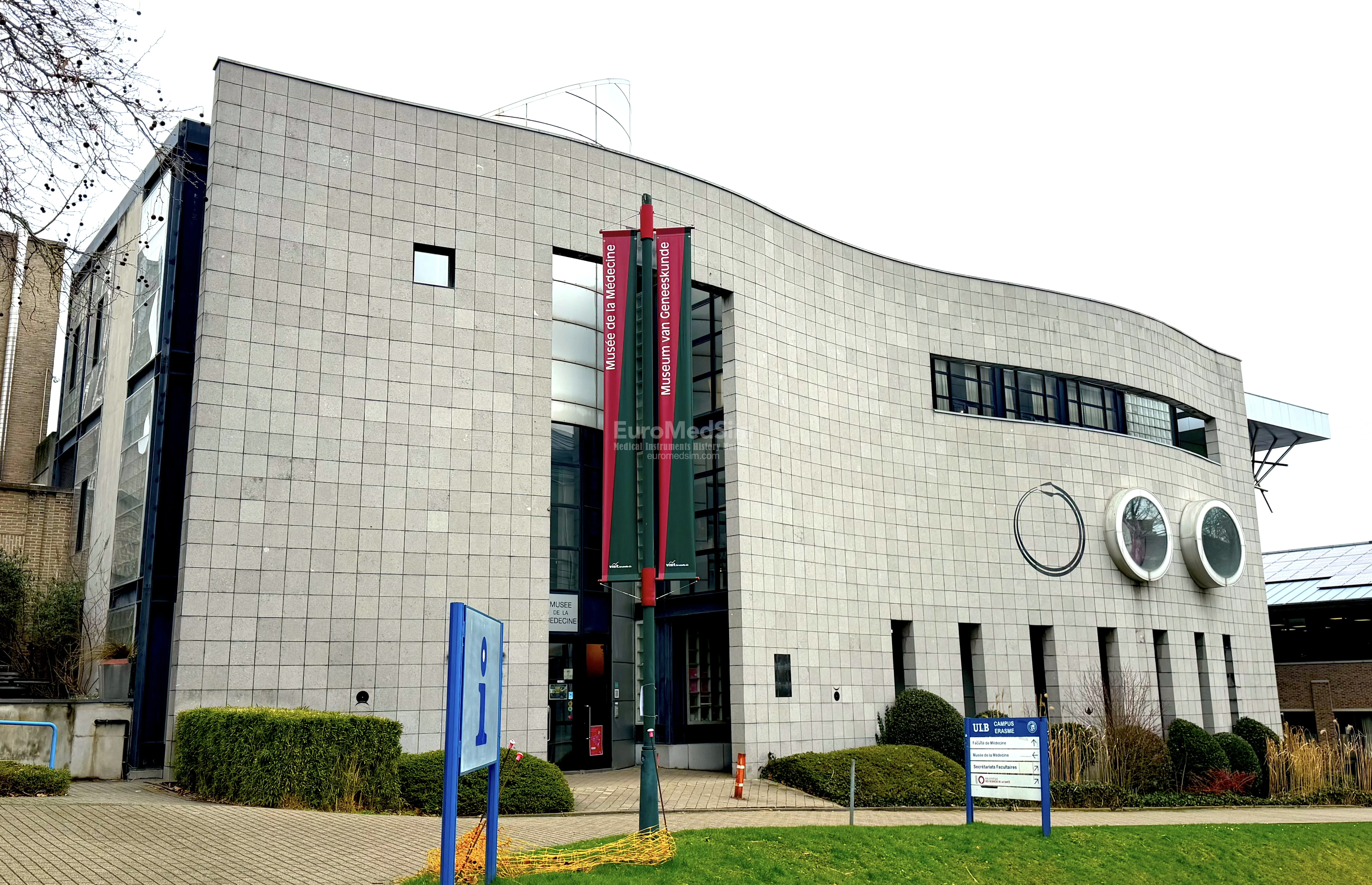


Museum of Medicine of Brussels
Museum of Medicine / Musée de la Médecine / Museum van Geneeskunde
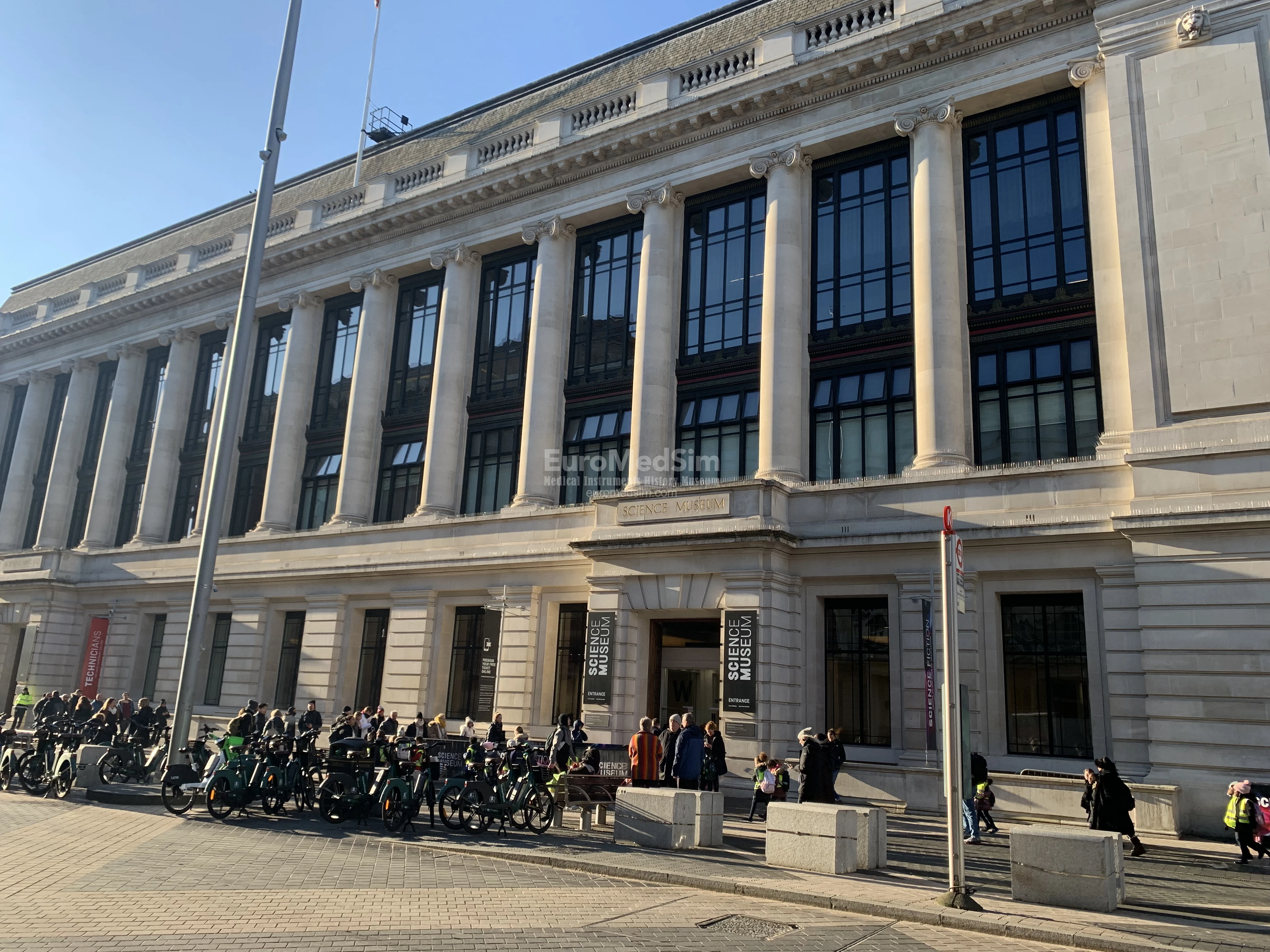
The Museum of Medicine (Musée de la Médecine / Museum van Geneeskunde) is located at the Campus Erasme of the Free University of Brussels (ULB — Université Libre de Bruxelles / Vrije Universiteit Brussel). The Museum opened its doors in 1994. Its exhibition is housed in a separate architecturally remarkable building next to the huge Erasmus Hospital complex in Anderlecht (Brussels-Capital Region).
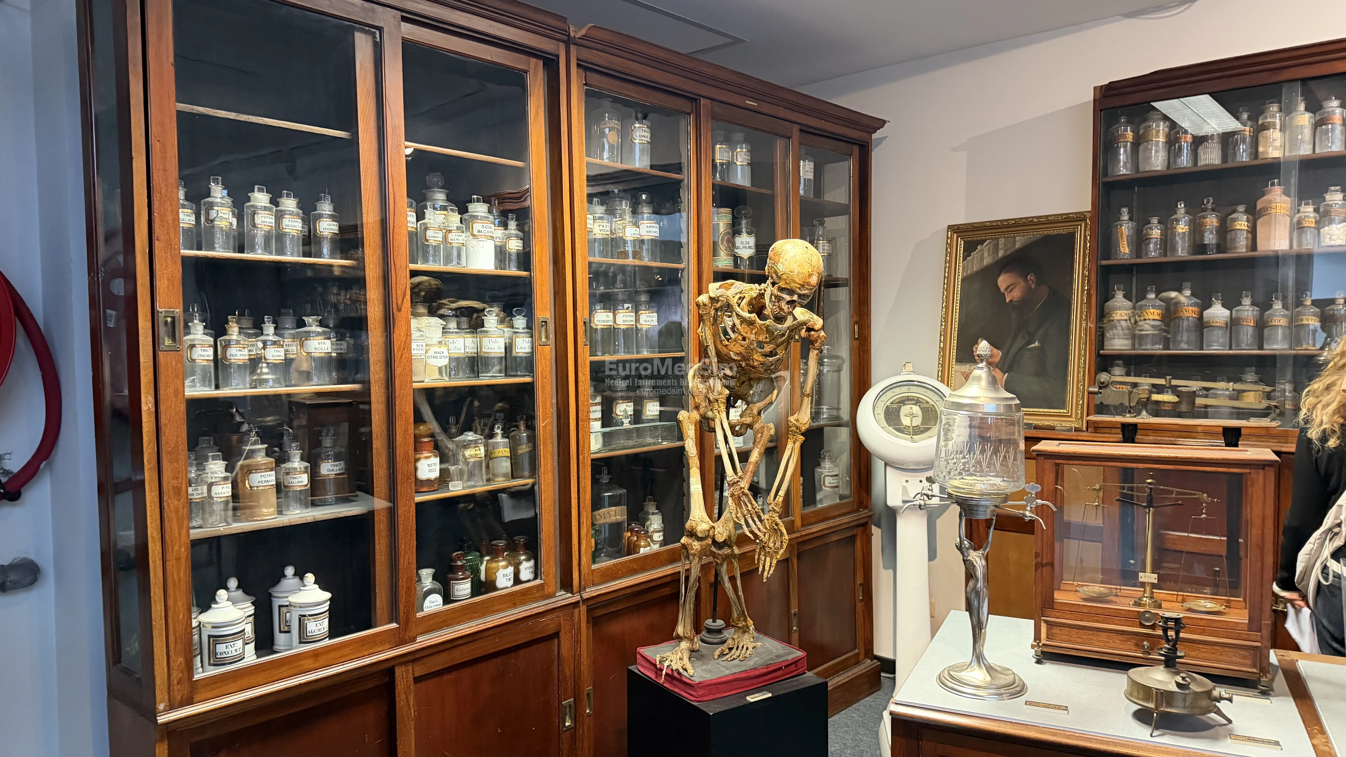
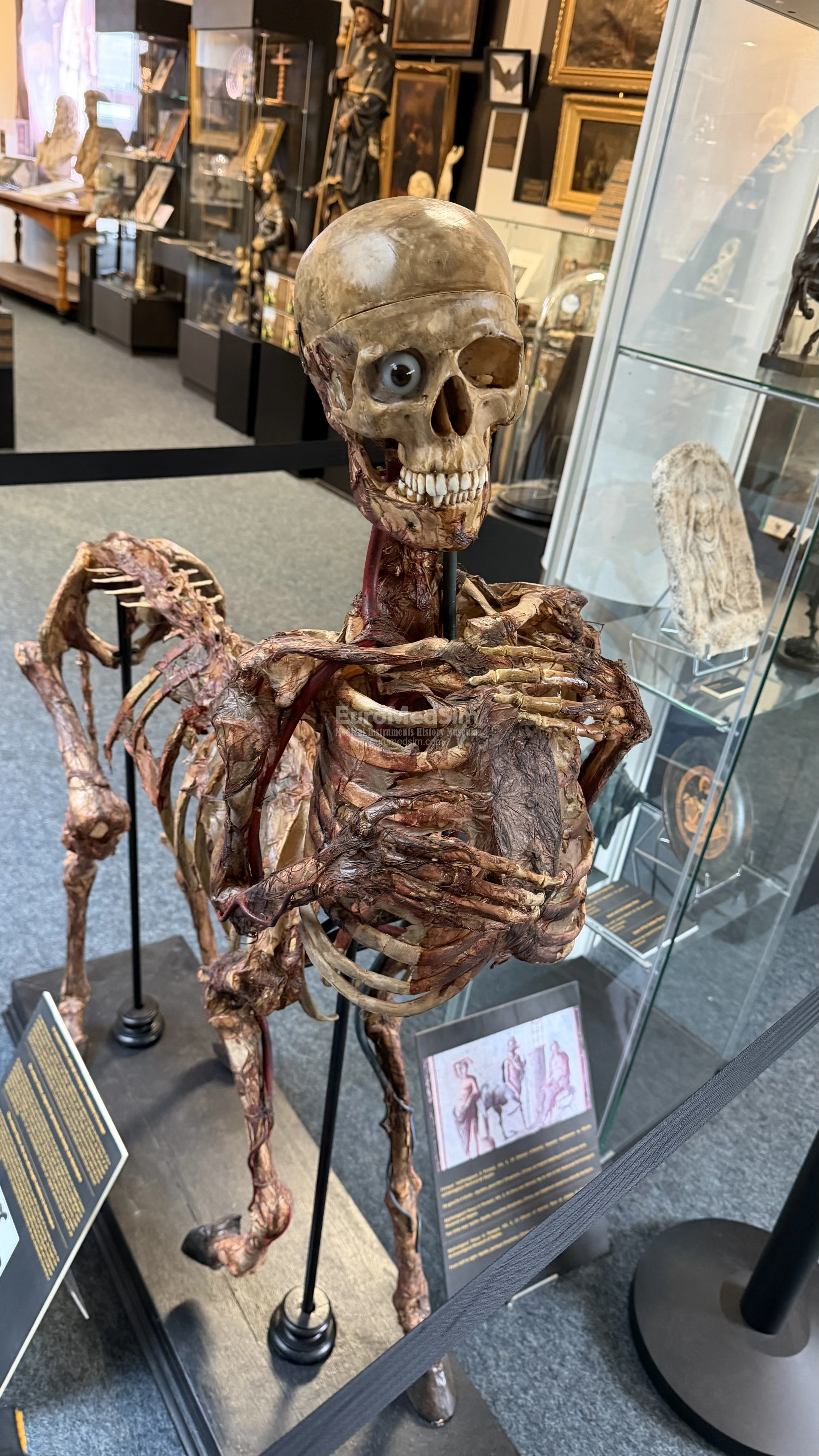
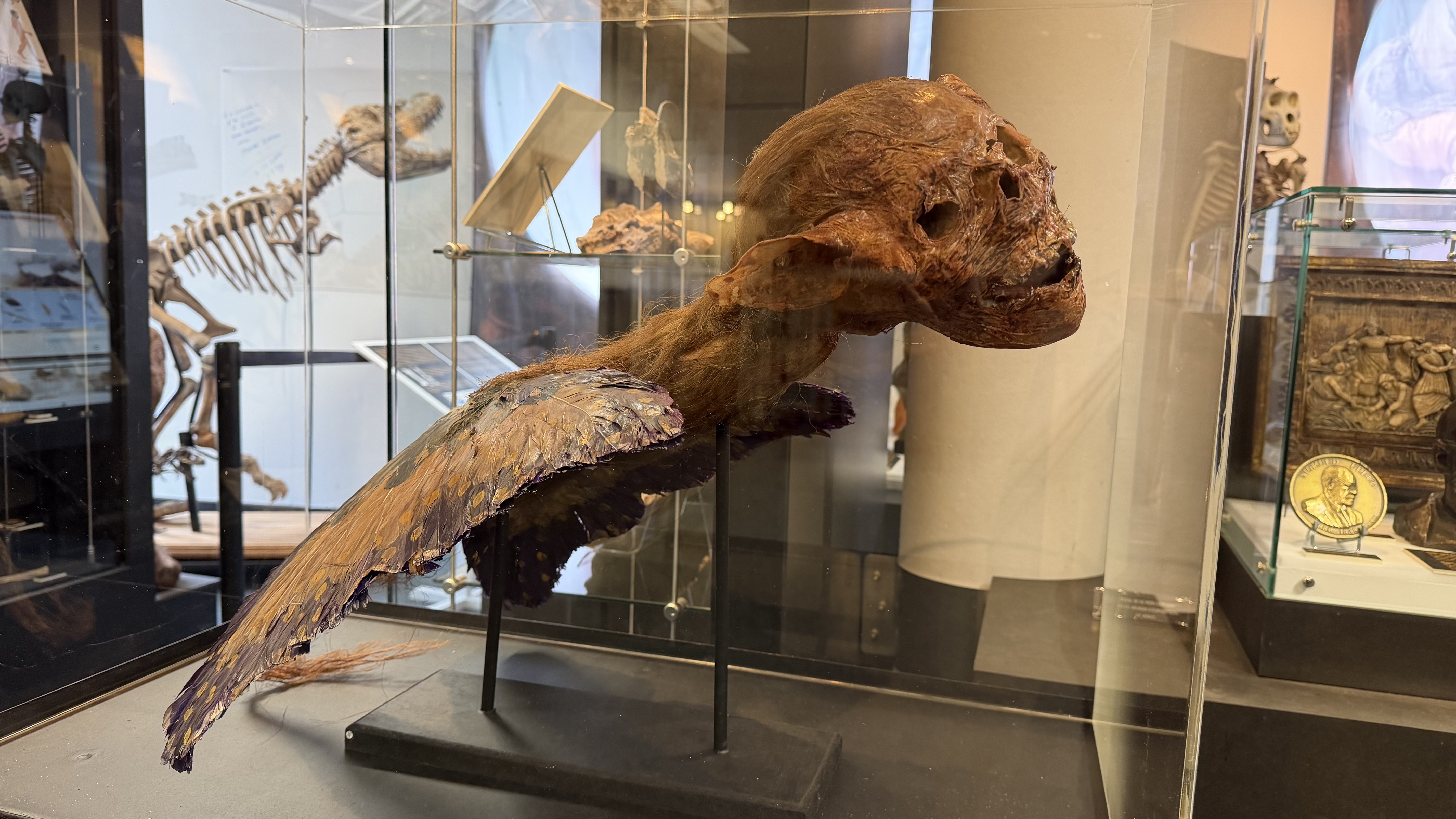
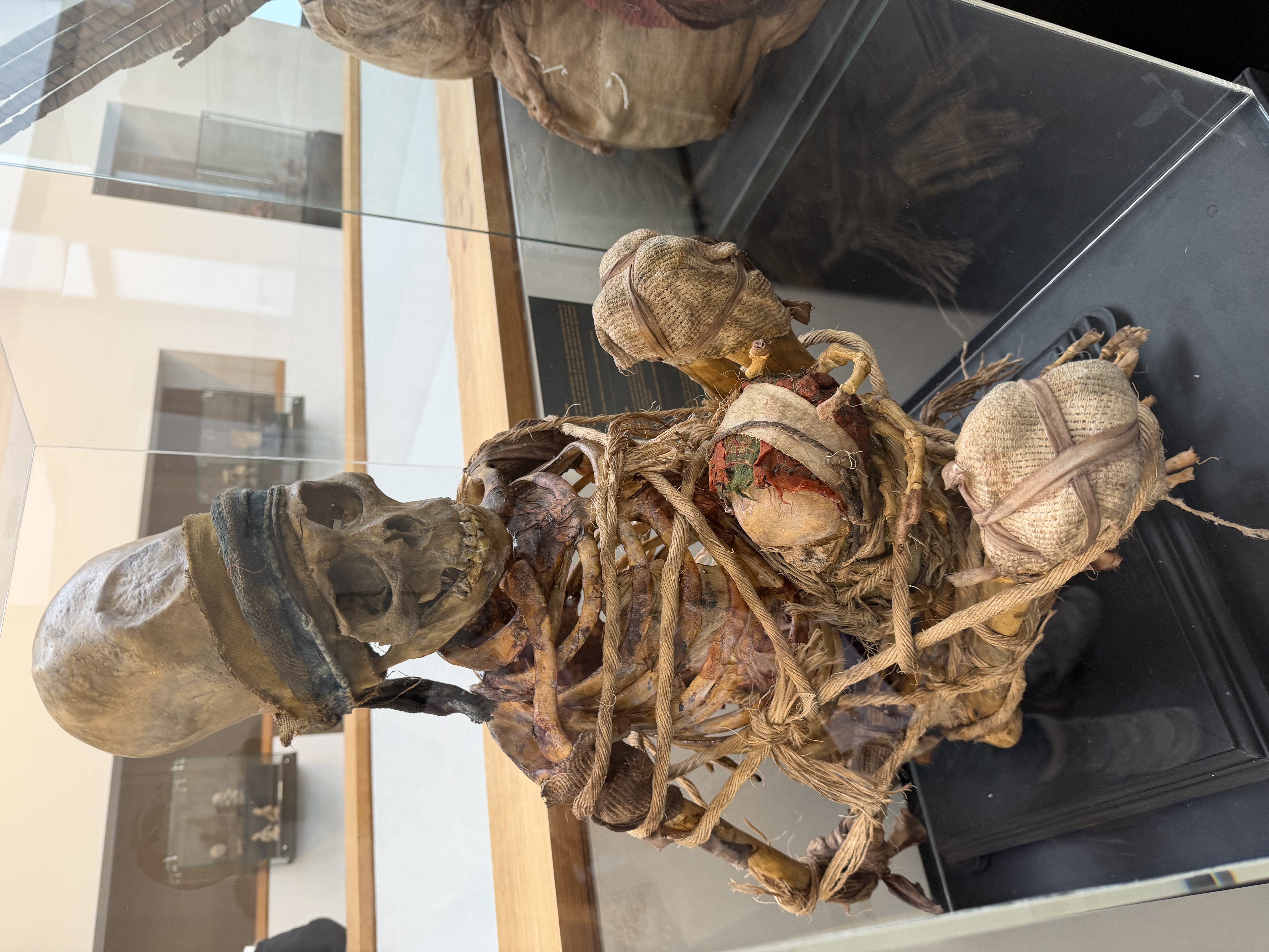
The museum offers a unique and sometimes fascinating journey through the history of medicine. Among its most unusual exhibits are figures of an alien, a centaur skeleton, and a harpy. Alongside these bizarre exhibits, the museum displays rare medical instruments, anatomical models, and historical artifacts that trace the evolution of medical knowledge.
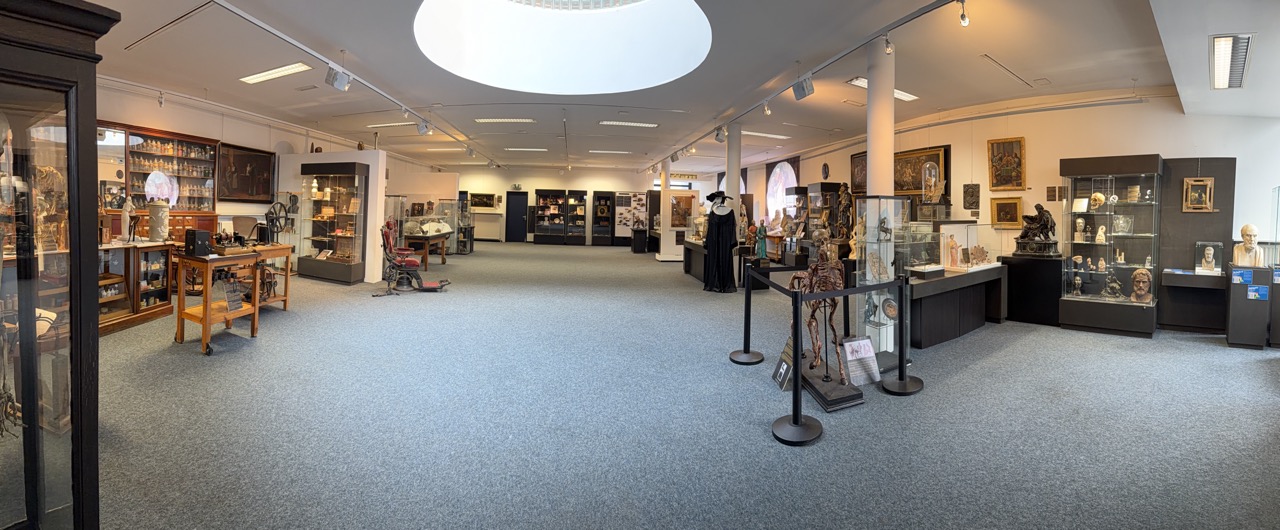
The exhibits are logically grouped into the following sections on four floors: Medicine in Egyptian Art; General History of Medicine (from Antiquity until the 20th Century); Pharmacy; Anatomical Waxes I (Anatomy, Obstetrics, Venereal Diseases); Medicine in Pre-Columbian Art. A distinctive feature of the museum is that many of the exhibits are displayed in a room without display cases, allowing visitors to examine them closely from any distance and from different angles, without glare or other distractions.
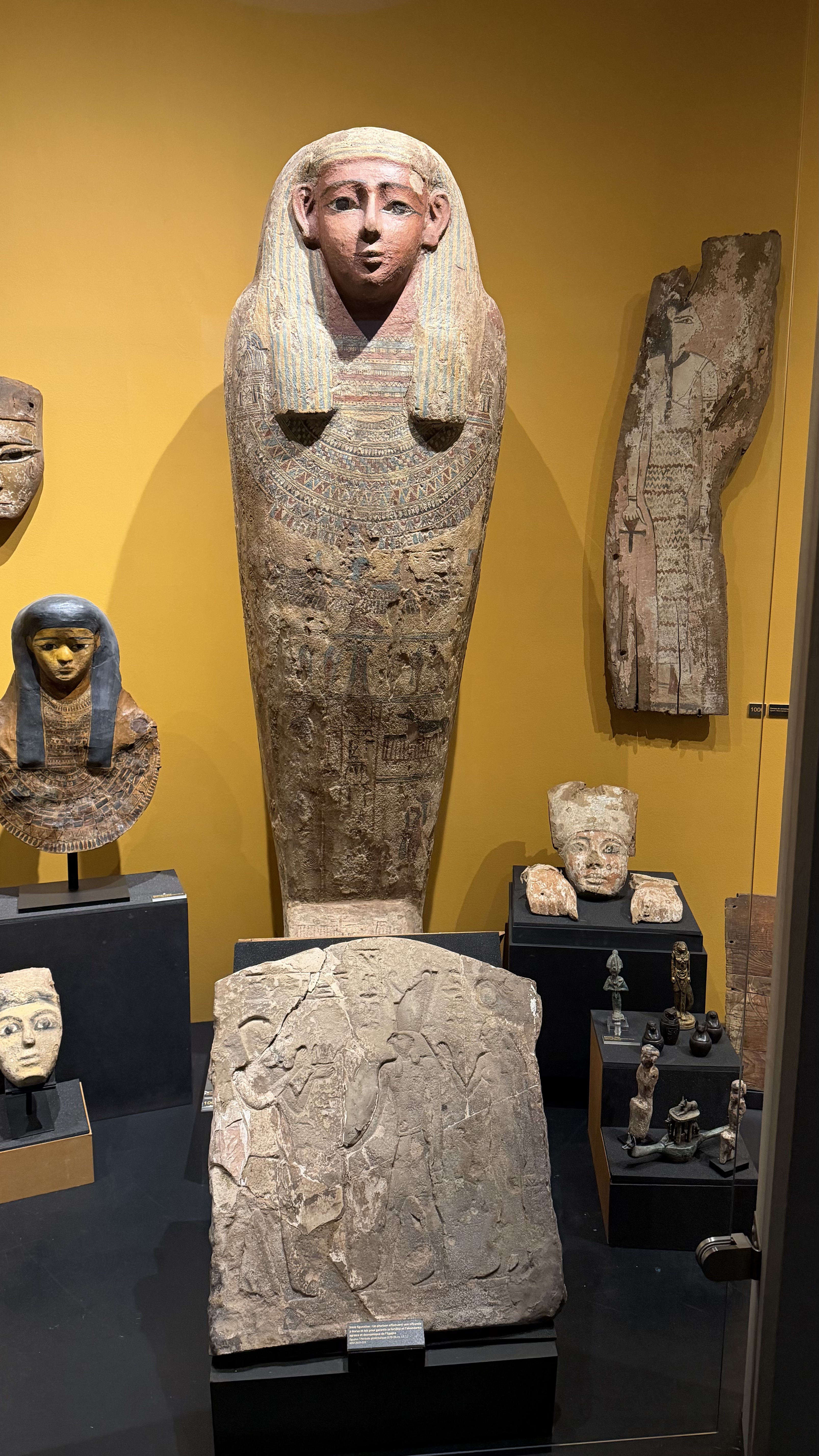
Underground Floor: Medicine in Ancient Egypt
In ancient Egypt, medicine was closely tied to religion and beliefs about the afterlife. The heart, called the "ib," was considered the seat of vital force, emotions, and desire. As the central organ, it sent the "metou" into the vessels—the stream that brought movement, thought, action, and speech.
Small cosmetic vessels and cream pots suggest the precious nature of their contents. A makeup palette made of schist in the form of an ibex was used to grind powders for eye makeup, which protected against sun and insects. The Egyptians used perfumed oils abundantly on their bodies and also coated statues of the gods with them, as perfume was considered an emanation of the divine. Oils were also used in the ritual of the opening of the mouth.
Ancient Egyptian religion believed in life after death, so mummification of the deceased was essential for survival in the next world. This practice continued into the Islamic period. The body was emptied of its organs, which were preserved in separate containers placed under the protection of the four sons of Horus. The body was then cleaned and dried with natron. After forty days, the natron was removed and the body was wrapped in bandages, with amulets placed between the layers.
A sarcophagus from the 25th–26th Dynasty (around 625 BC, Late Period) features thin eyebrows, and the pectoral extends to the shoulders with two falcon heads crowned by solar discs. Lower down, Isis spreads her wings while the four sons of Horus surround an offering table with three loaves. Above on the right, Anubis the jackal-god appears with Thoth as a baboon in the solar disc. These illustrations refer to the ritual directed by Anubis, who brings the deceased by the hand before the scales where the heart is weighed against the feather of truth and justice. If the heart is not lighter than the feather, death is final.
The Egyptians believed in natural and supernatural causes of illness. They called on priests and incantations and wore amulets, which were also placed on mummies for protection. These amulets invoked divinities and expelled demons while remedies were applied. Horus, the falcon-god, son of Isis and Osiris, who won the battle with his uncle Set and received the throne of Egypt as his prize, is often represented as an adolescent with hair to one side, finger in his mouth, and a prominent phallus, ready to combat diseases sent by his mother and Set. His mother Isis, who nursed him, reminded him that she had the power to decide life and death. She possessed extremely powerful magical gifts, especially as a protective mother while nursing.
Main Hall at the 1st Floor
To the left of the entrance to the main hall, visitors are greeted by a 200-year-old English pharmacy. Cabinet contains 274 flasks: plant extracts (eucalyptus, valerian, quinquina, foxglove); metal preparations (iron, zinc, bromide); semi-purified substances (opium, colchicine) presented in the form of liqueurs, powders, pastilles, aqueous solutions, acids, oils, tinctures, gums, wines, and so on. This arsenal had developed thanks to the introduction of measurement and laboratory research since the previous century, particularly with Lavoisier. Nevertheless, these preparations were relatively ineffective. One had to wait until the second half of the 19th century for pharmacy to base itself on extractive chemistry and synthesis. Scheele isolated certain organic acids from plant drugs but never any base; the opium alkaloid would be one of the first extracts, followed by emetine from ipecacuanha and cinchonine from quinquina.
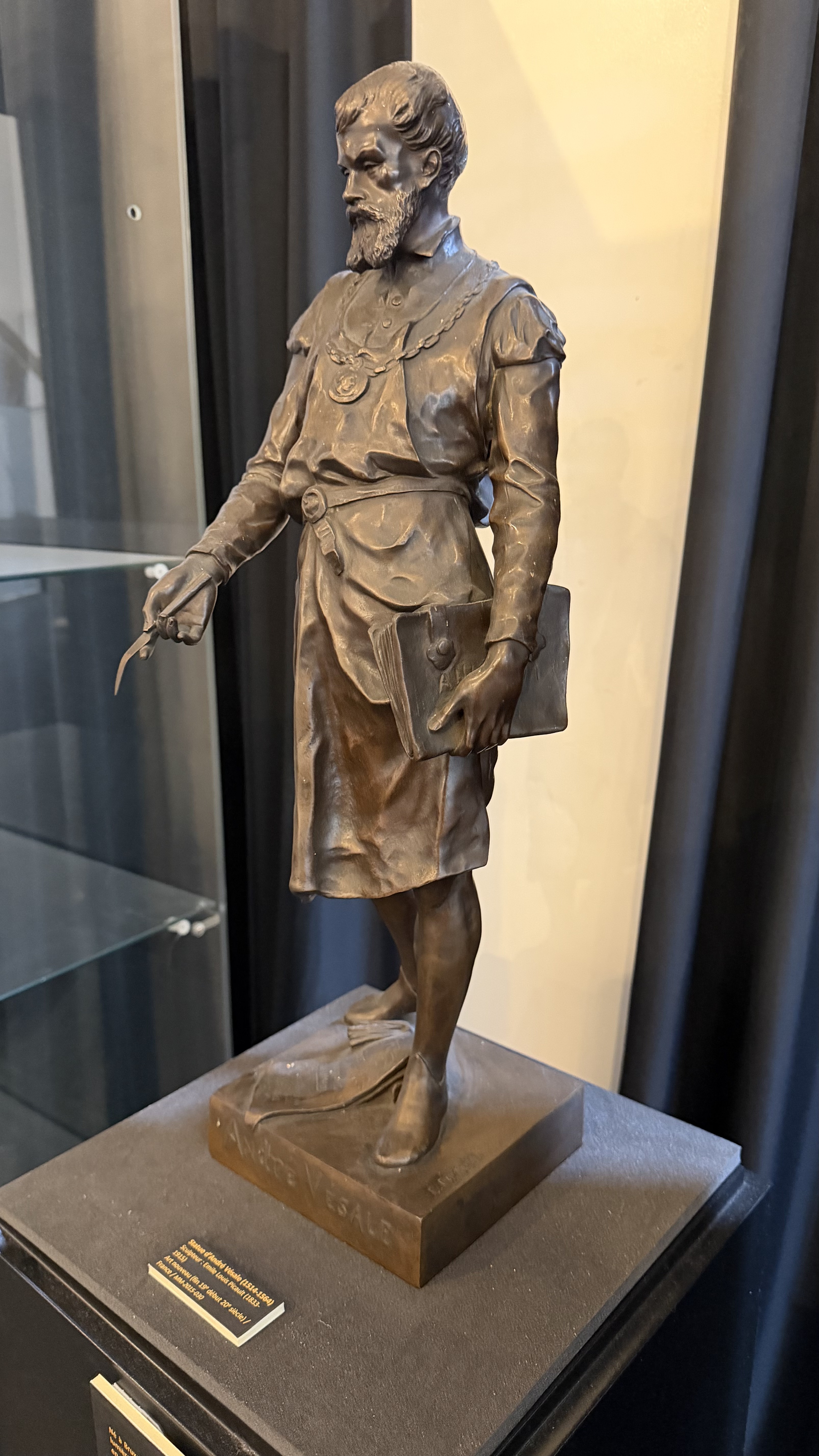
Andreas Vesalius (whose bronze sculpture is exhibited here) was born in Brussels on December 31, 1514. Born into a family of physicians, he began his studies at the Trilingual College in Louvain, then at the Paris Medical College. He recovered the bodies of hanged criminals and dissected them, then began writing textbooks, including the famous "De Humani Corporis Fabrica" in 1543. This book is the foundation of anatomy. The skeleton, muscle attachments, nerve pathways, and various organs of the human body are described with precision, correcting Galen's errors. The illustrations in this great work are by Calcar, a pupil of Titian.
The invention of firearms made battlefield injuries much more serious and the treatment of the wounded more complex. The doctor tried to put the patient to sleep with mulled wine or opium; the patient was "woken up" by inhaling vinegar. Ambroise Paré (another fantastic bronze figure), born in 1510, replaced the cauterization of wounds and the application of boiling oil by cleaning with rose water. He improved techniques for ligating blood vessels, used probes to find the trajectory of projectiles and removed them while limiting surgical intervention. He owes his immense popularity not only to his skill as a surgeon, but also to his talent as a writer. The numerous books he wrote in French (rather than Latin), accompanied by numerous illustrations, made him famous throughout France, and his Oeuvres, a complete collection of his works published in 1575, were translated into the main European languages and reprinted numerous times.
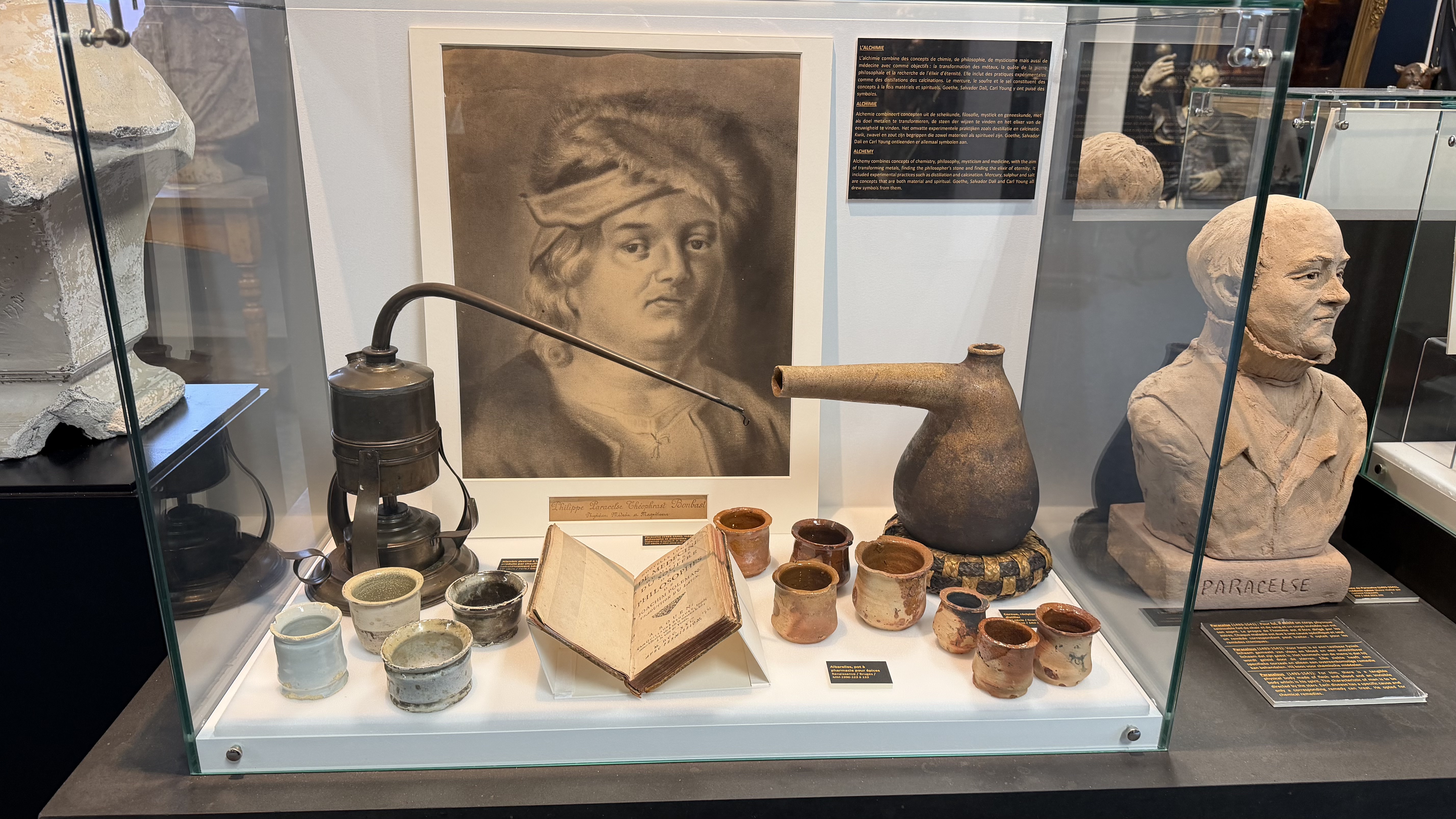
Until the end of the 19th century, remedies were derived from the animal, vegetable, and even mineral worlds, under the influence of Paracelsus and the Brussels-born Van Helmont. Medicinal plants were stored dry, hung from the ceiling, or kept in labeled drawers, often decorated. Mixtures were prepared in pill boxes, apothecary pots, mortars and pestles, ointment pots, and theriac jars. Stills were used for the preparation of alcohols, tinctures, essences, and aromatic waters. Almost all substances used were prepared in vinegar, sugar, or alcohol. There were also secret remedies.
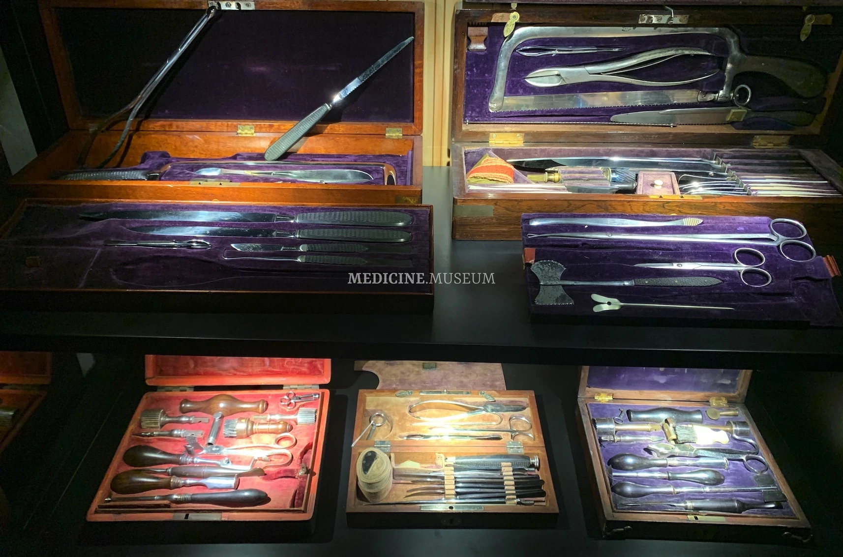
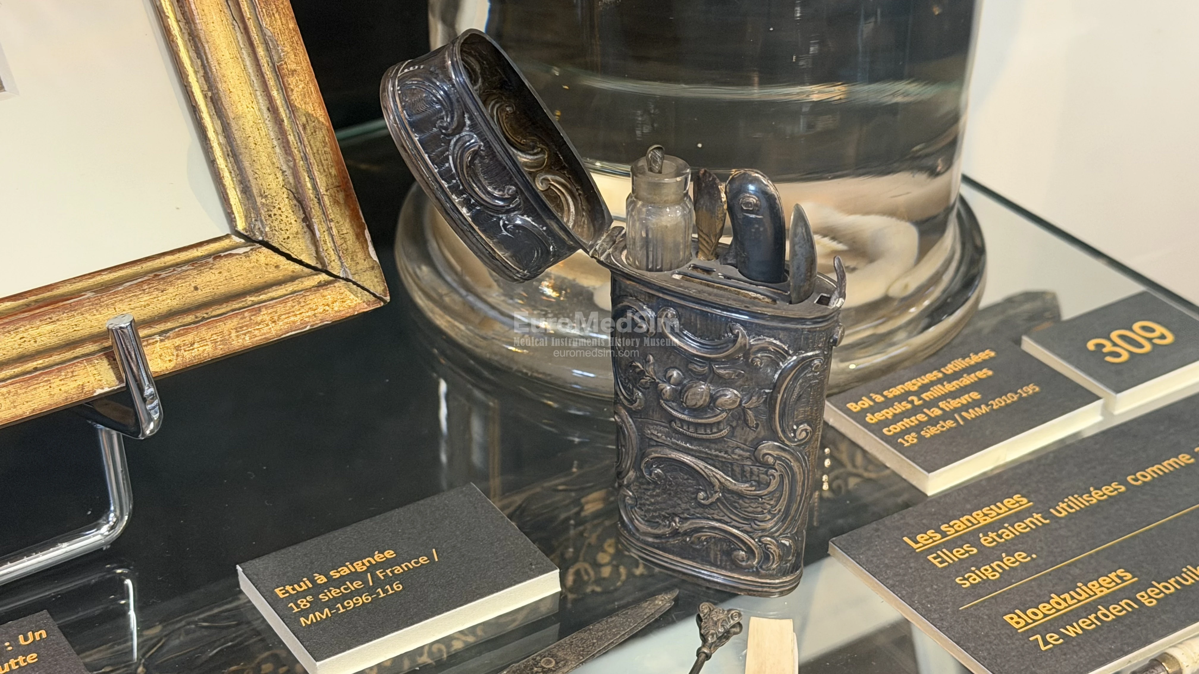
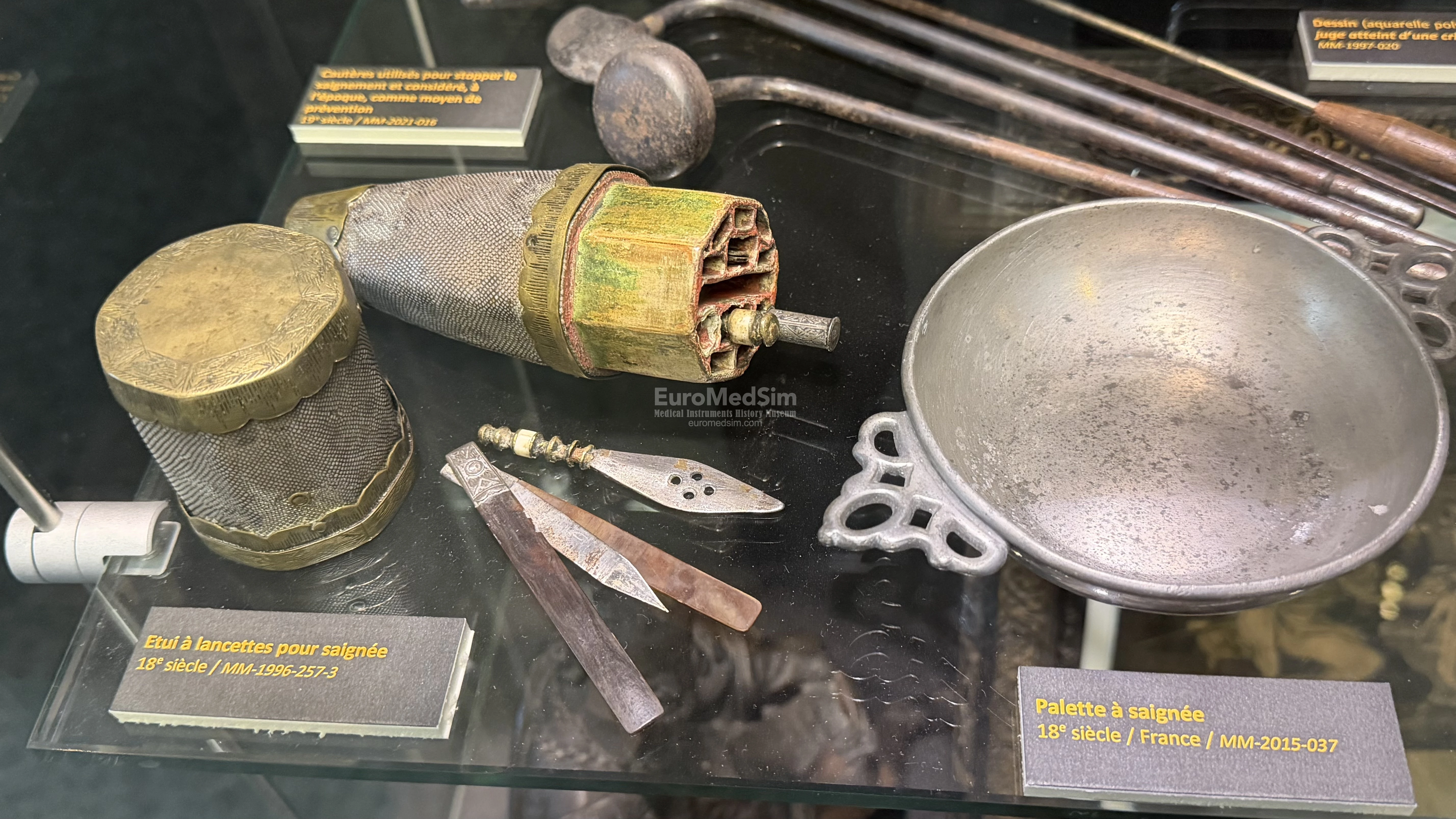
Bloodletting also existed in ancient Egypt, where it was observed that sick hippopotamuses bled themselves by rubbing against reeds in the Nile before rolling in the mud. In Antiquity, bloodletting was used to restore the balance of the four humors. In the Middle Ages, bloodletting was supposed to lighten the eyes, calm the brain, sharpen the wits, warm the bones, and increase strength. It was mainly used on the obese and impetuous to allow them to resume the pleasures of the table, and on monks to favor chastity. It became a cure-all in the 17th century, performed 8 to 20 times. It was considered that blood, particularly its excess, was responsible for illness. The odor of blood also gave information about its quality. The physician had sharp lances and an indented palette to collect blood and water, along with vinegar or rosemary wine in case of fainting.
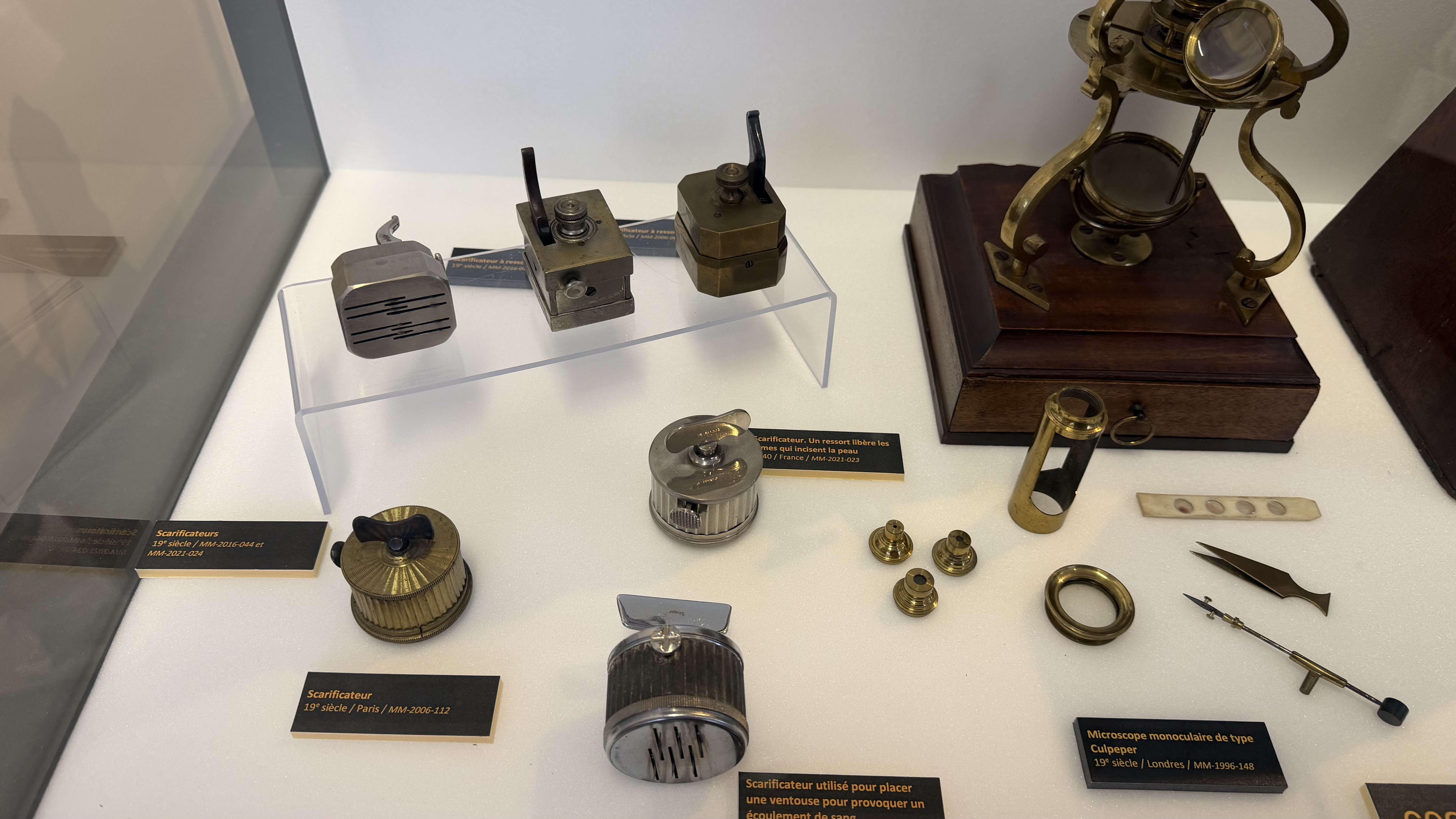
In the 19th century, bloodletting with blades in a square or octagonal box also had popularity. On pressing the switch, several blades positioned to cut the skin. Bloodletting was carried out on the forearm, but also on the temples, the base of the tongue, and the jugulars. It was also used by Jenner to apply cowpox pus for vaccination.
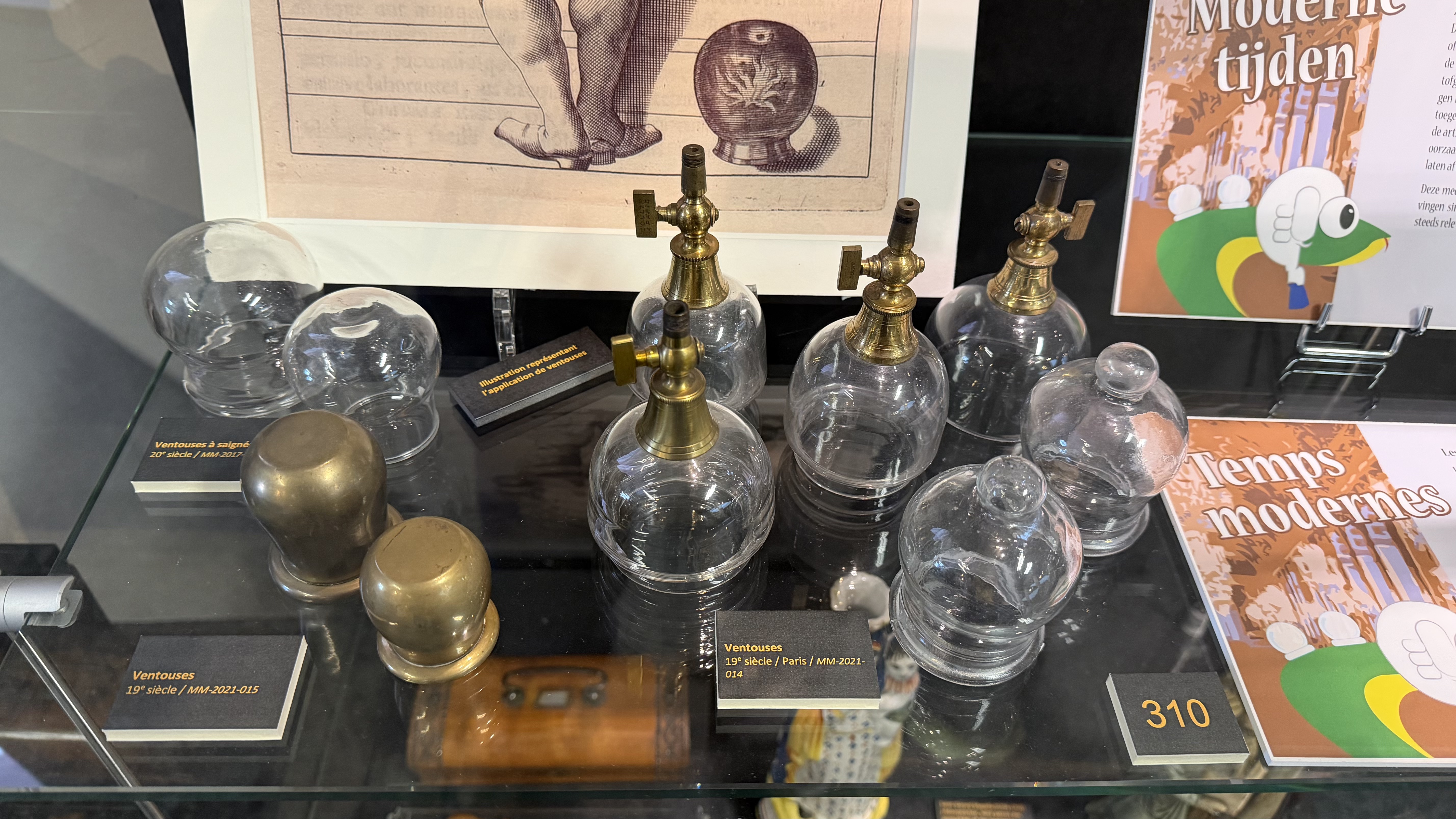
The vacuum created by cupping glasses opens the pores and superficial blood vessels. It was supposed to create decongestion near the affected organ, extracting humors and excess blood. They were applied under the nipples to contain menstrual bleeding, to the thighs to stimulate it, over the kidneys to move stones into the bladder, and so on.
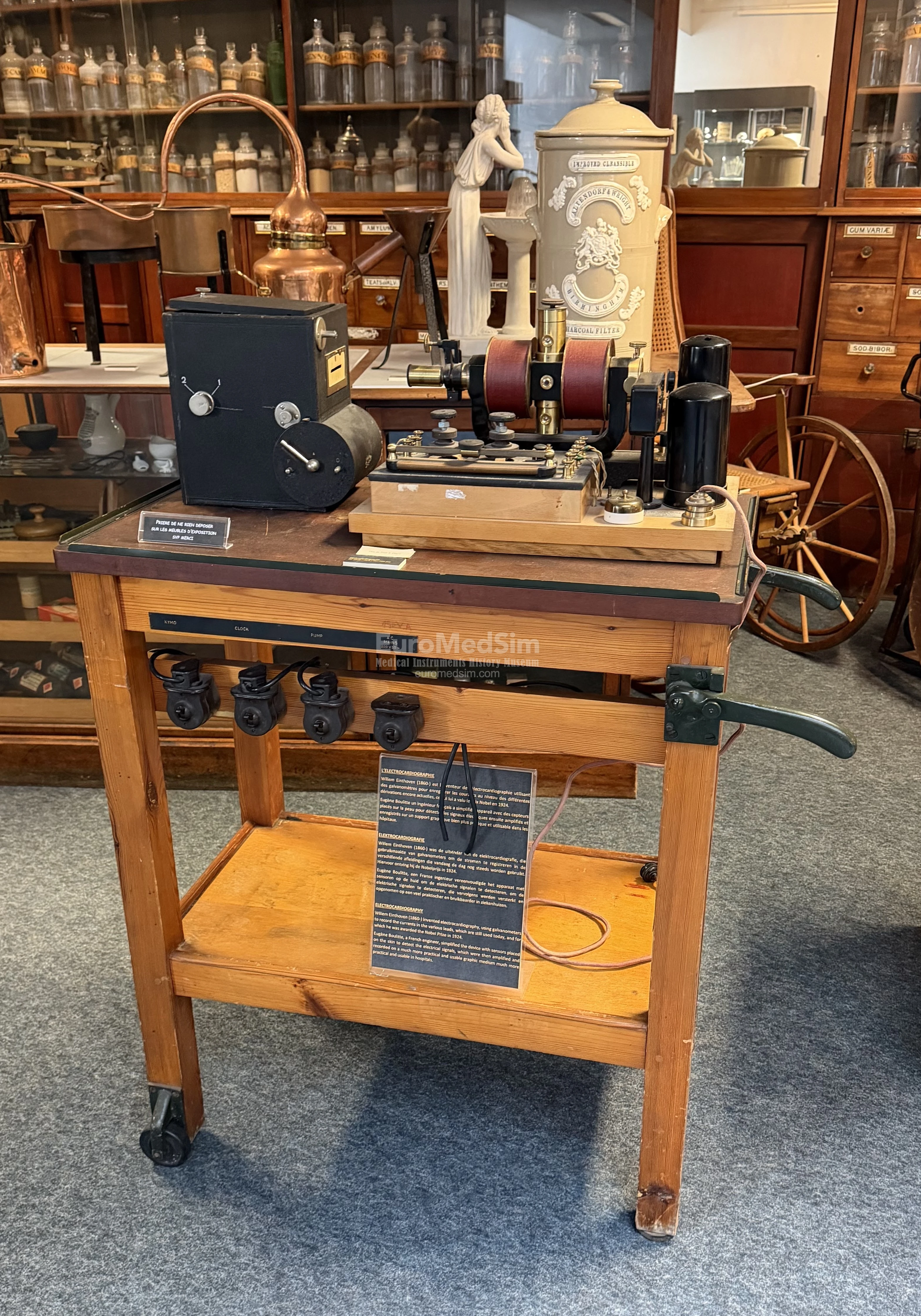
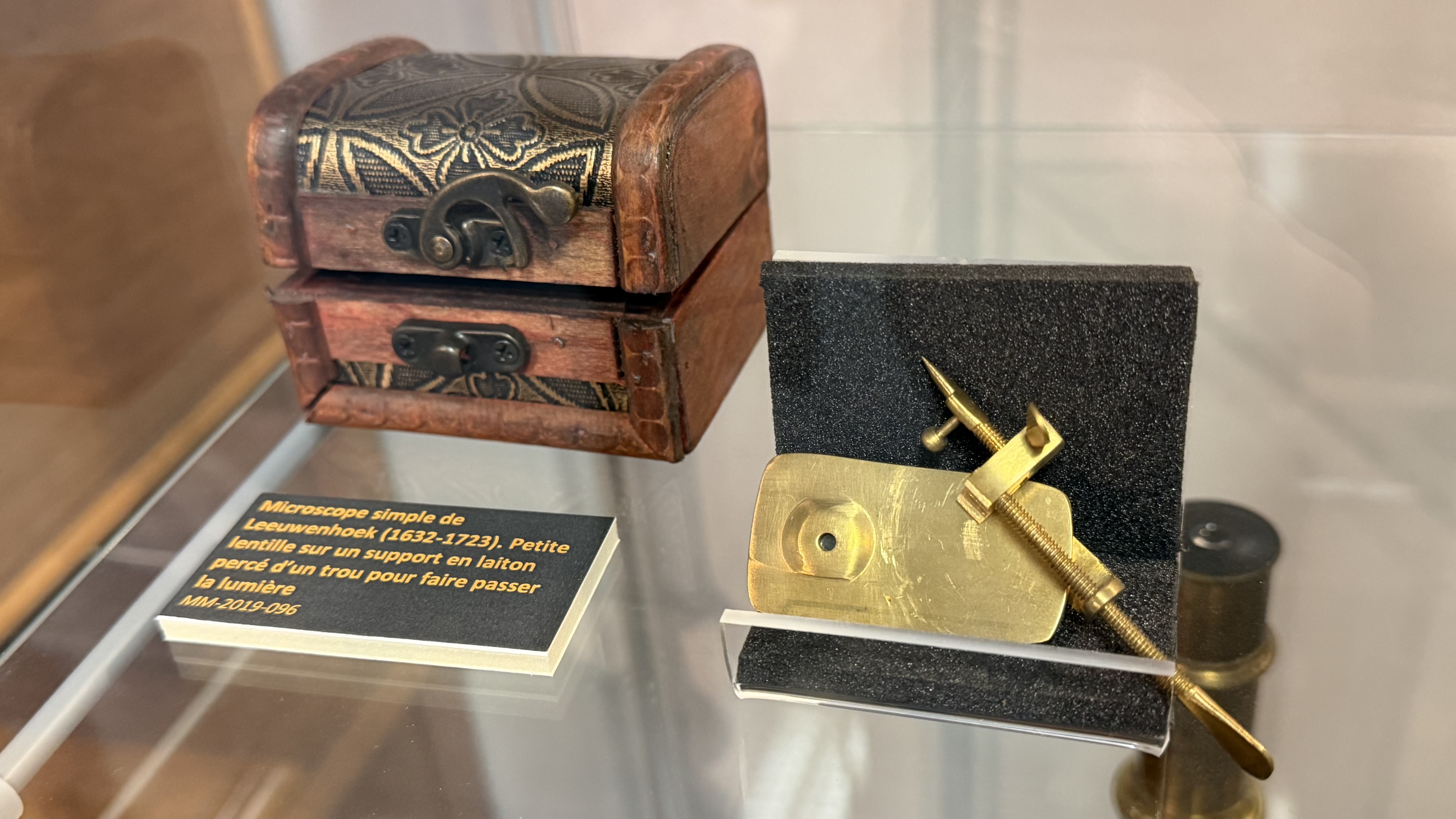
Born in 1632, Leeuwenhoek began by mounting biconvex lenses between two rectangular plates with holes for magnification — thus the first microscope has been invented. In this way, he could identify red blood cells, white blood cells, capillaries, the contents of semen, and so on. But his technique was discredited because it produced many artifacts, to the point that Bichat considered microscopic analysis a source of errors. One had to wait until 1820–1840 for the invention of achromatic lenses made of several different lenses welded together, which allowed avoidance of distortions. At the same time, Carl Zeiss introduced condensers and Jan Purkinje developed microtomes.
Jules Bordet graduated in medicine from the Free University of Brussels in 1892 and won the Nobel Prize in Medicine in 1919. Previously, he had worked at the Pasteur Institute on phagocytosis of white blood cells and the destructive power of specific antibodies against bacteria. He isolated the whooping cough bacterium and developed a test to detect syphilis, known as the Bordet–Wassermann reaction.
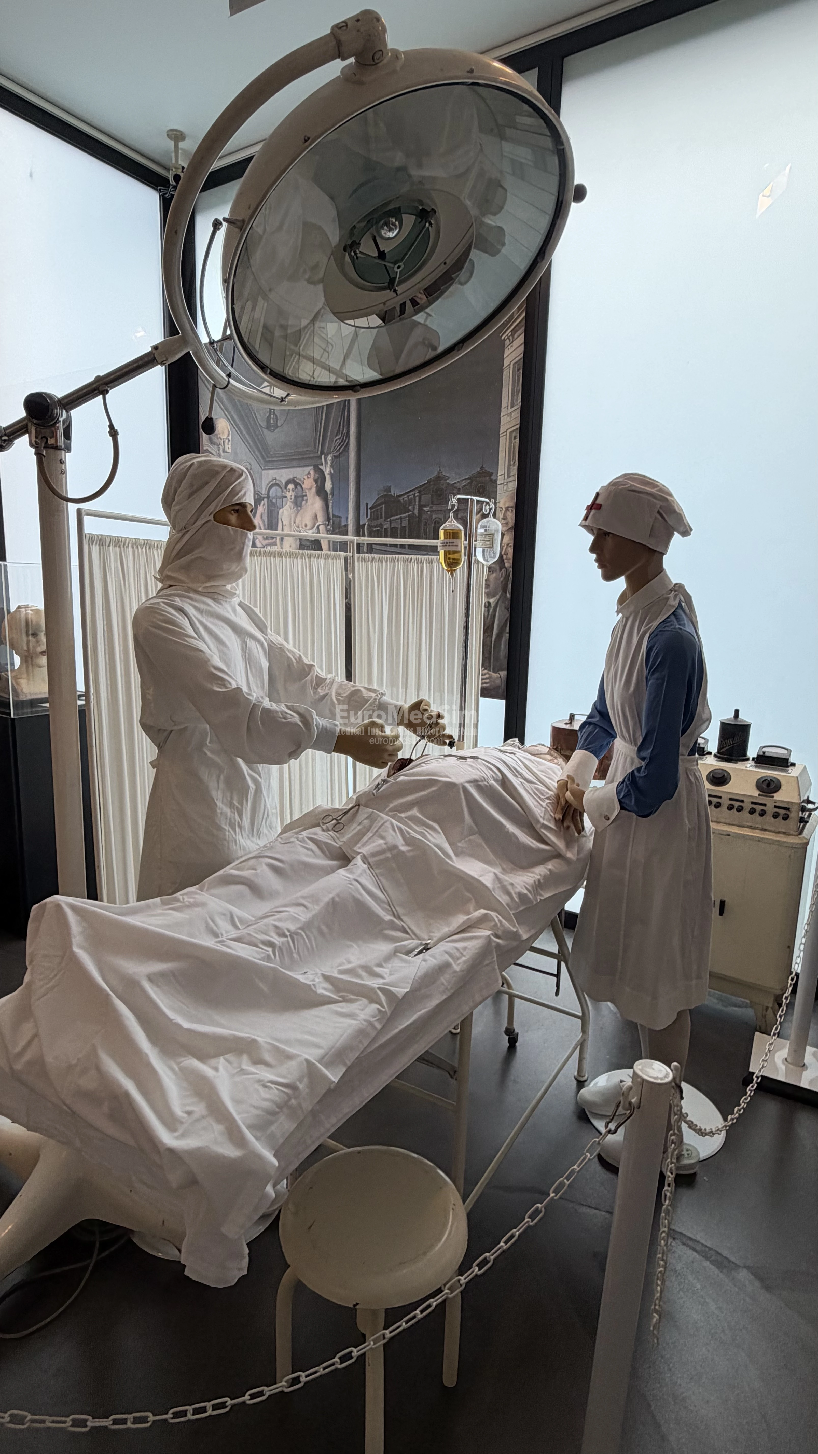
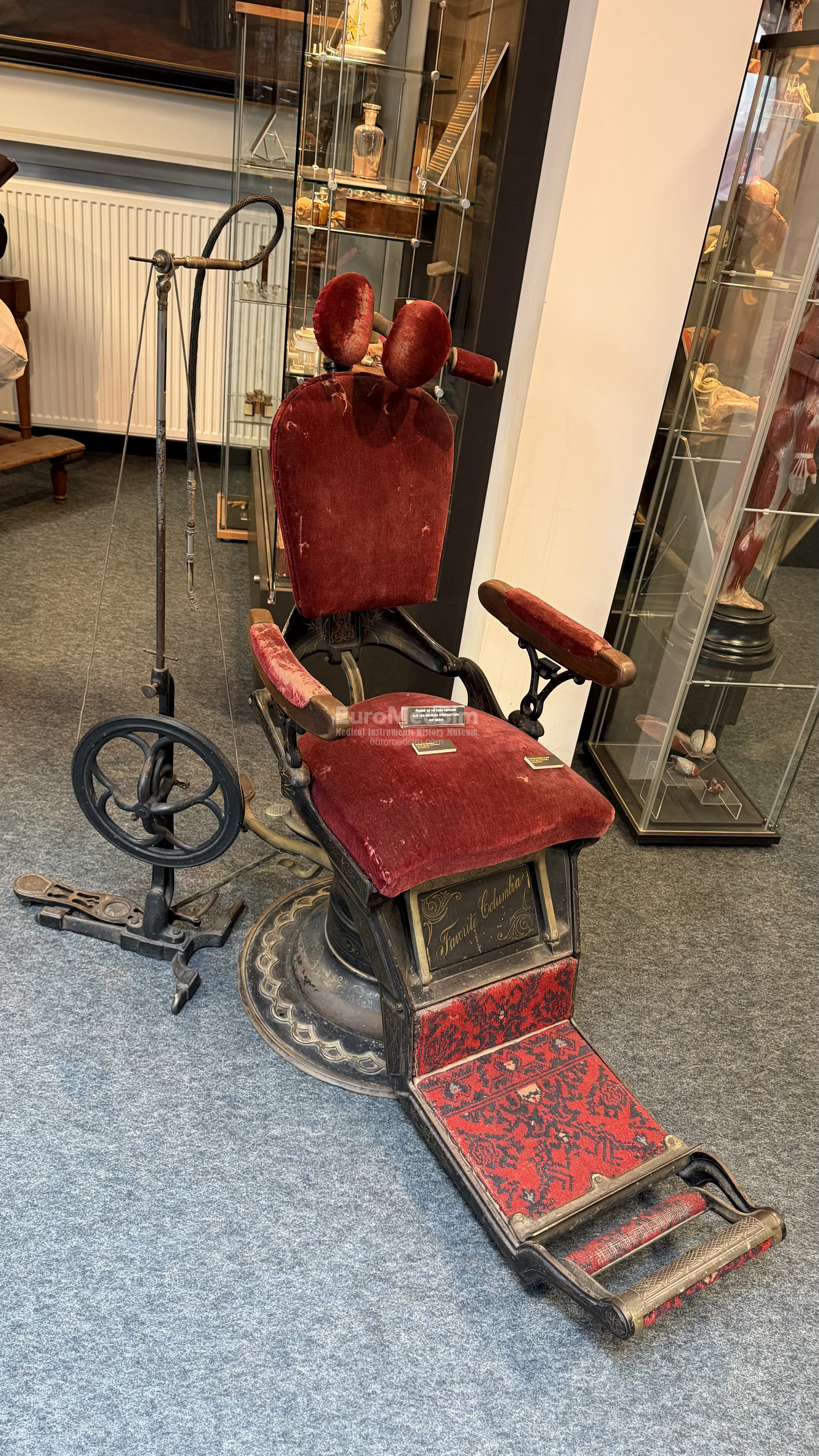
Surgery Room: 1st Floor
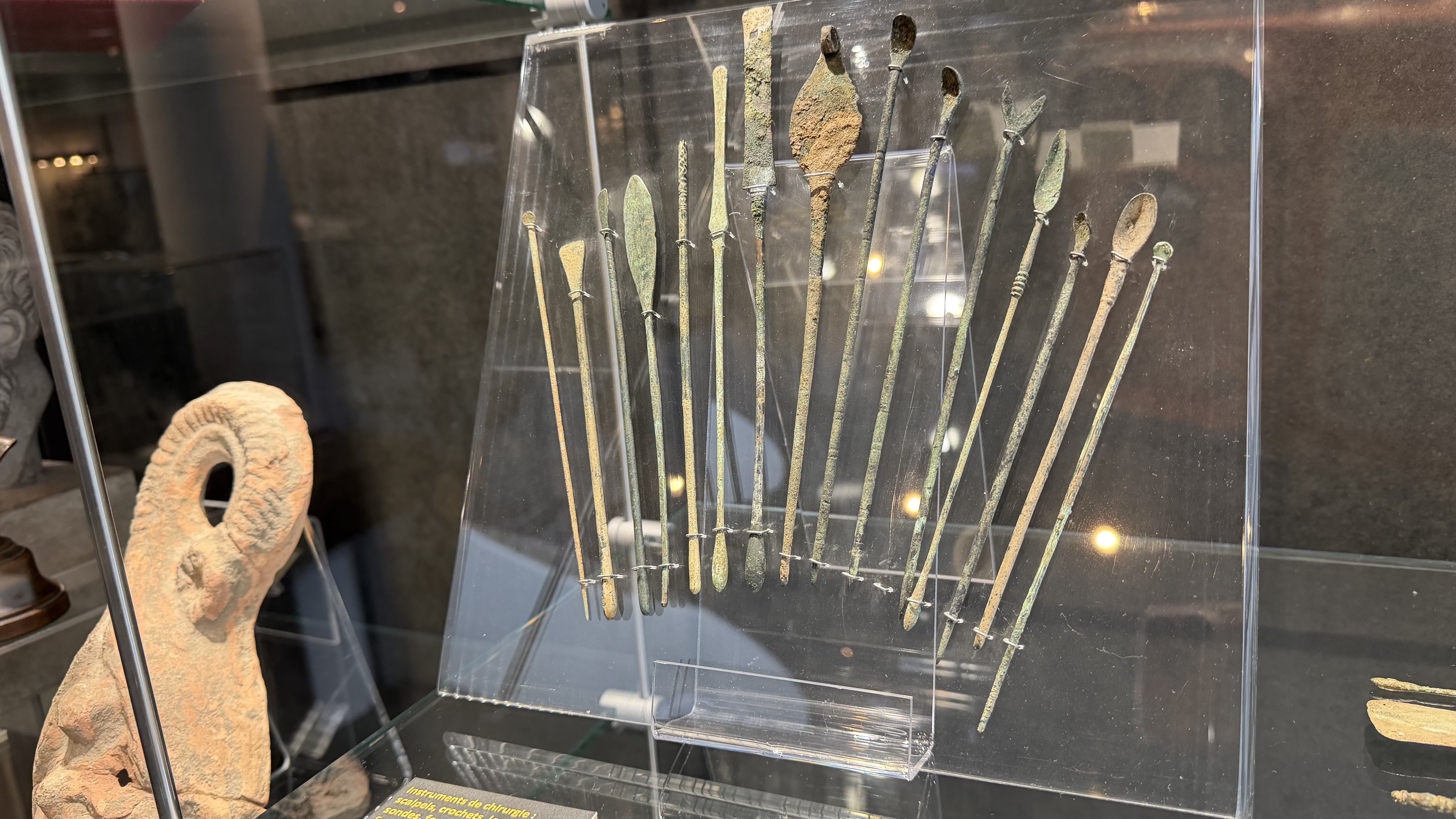
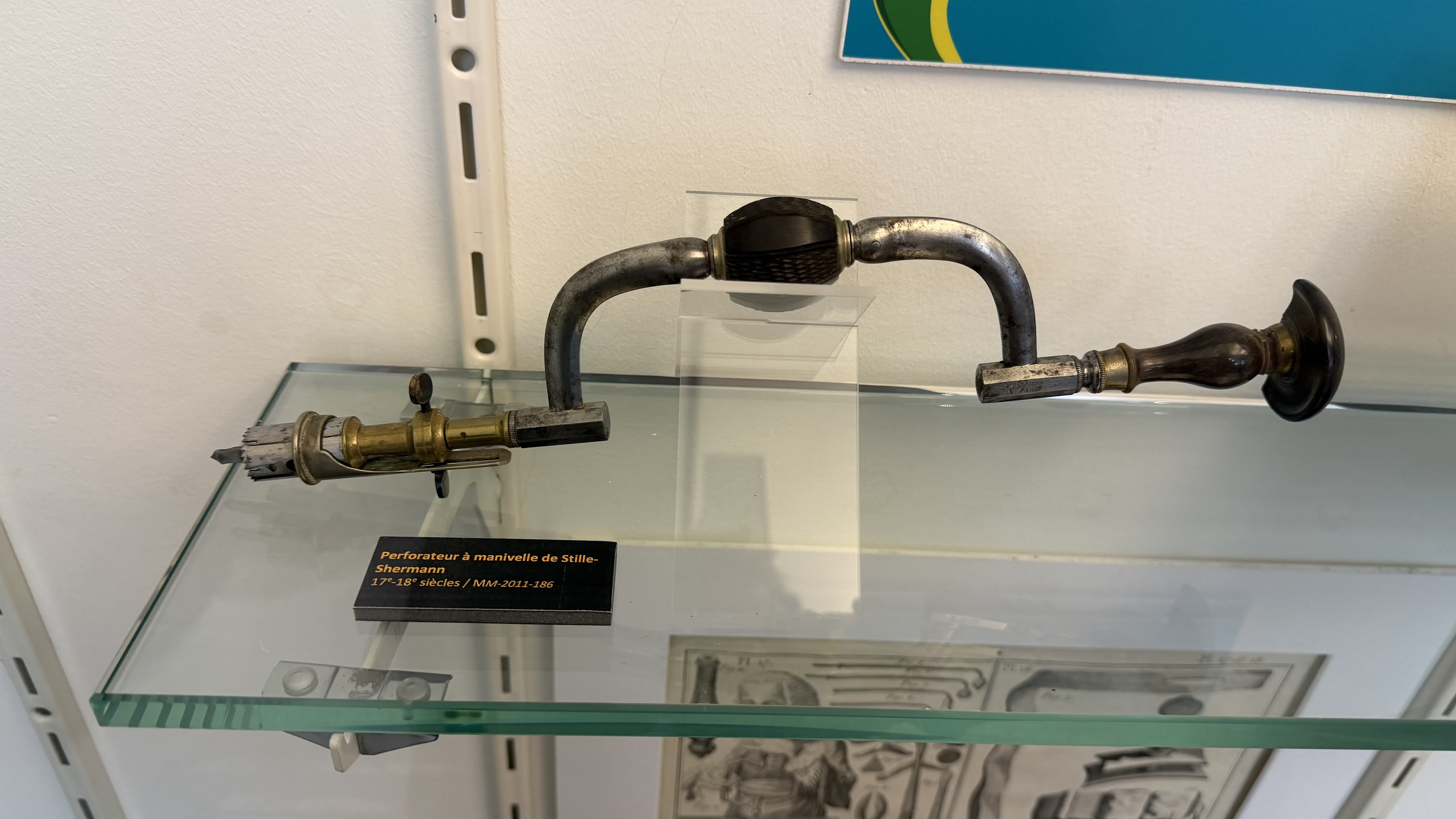
The term "surgery" is derived from the Greek words "cheir" (hand) and "ergon" (work). In Latin, "chirurgia" signifies the treatment of wounds and deformations by the intervention of hands and the use of instruments. The first indications are of minor surgery: treating wounds, repairing battle injuries, incising abscesses, performing trepanations, fixing fractures. This was made possible by rudimentary instruments and knowledge gained by Neolithic hunters from wounds they inflicted on their prey. Homer's Iliad, written around 800 BC, reports that doctors had developed expertise in treating war wounds. They easily treated superficial wounds, notably those from arrows; those from javelins and swords were more difficult. In 460 BC, the Hippocratic corpus described with precision techniques for reducing fractures, trepanation, shoulder luxation, treating superficial wounds, opening the chest to evacuate pus, and even inserting drains using variety of bronze instruments. To relieve pain, they used mandrake and sponges soaked in opium.
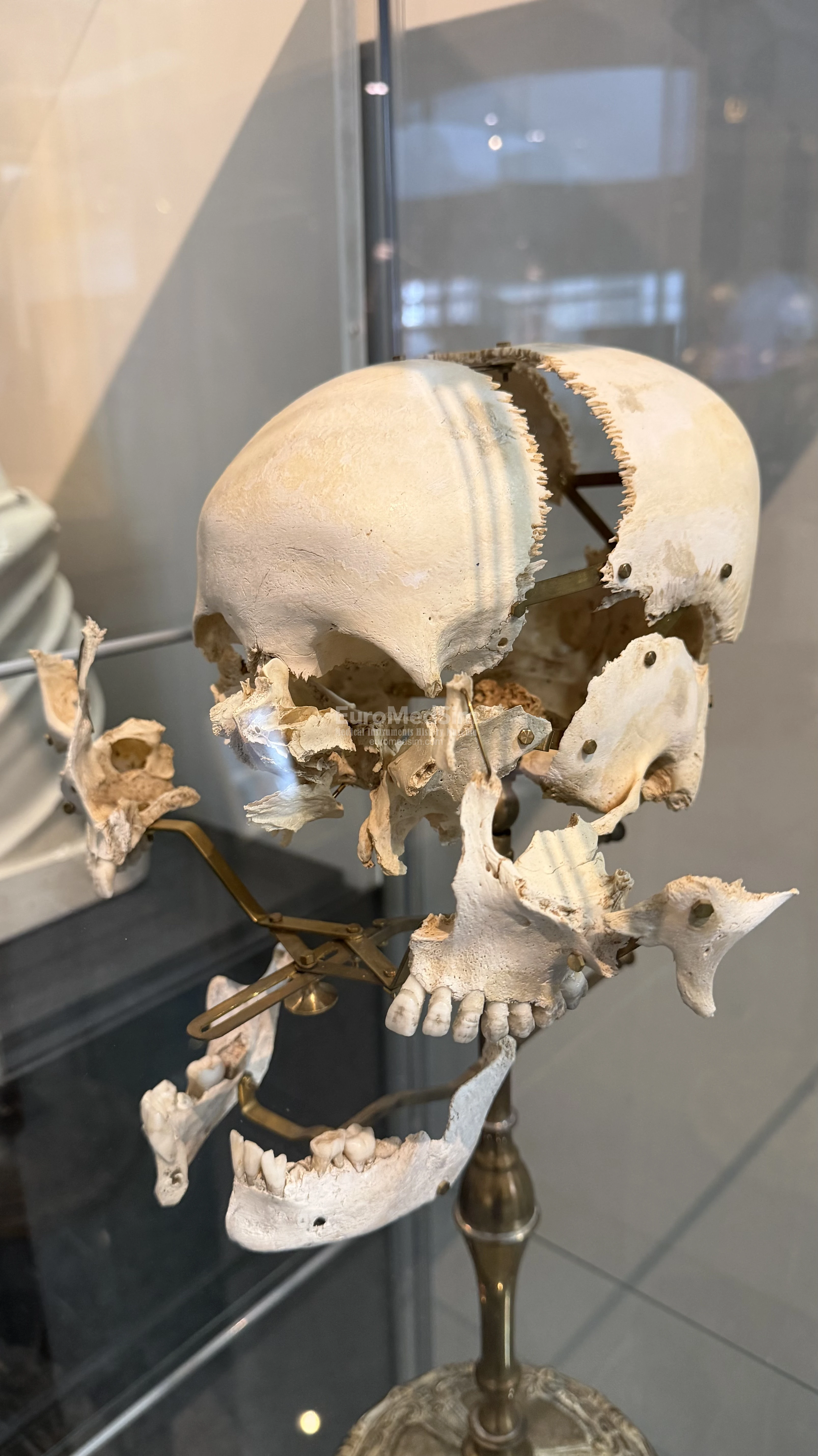
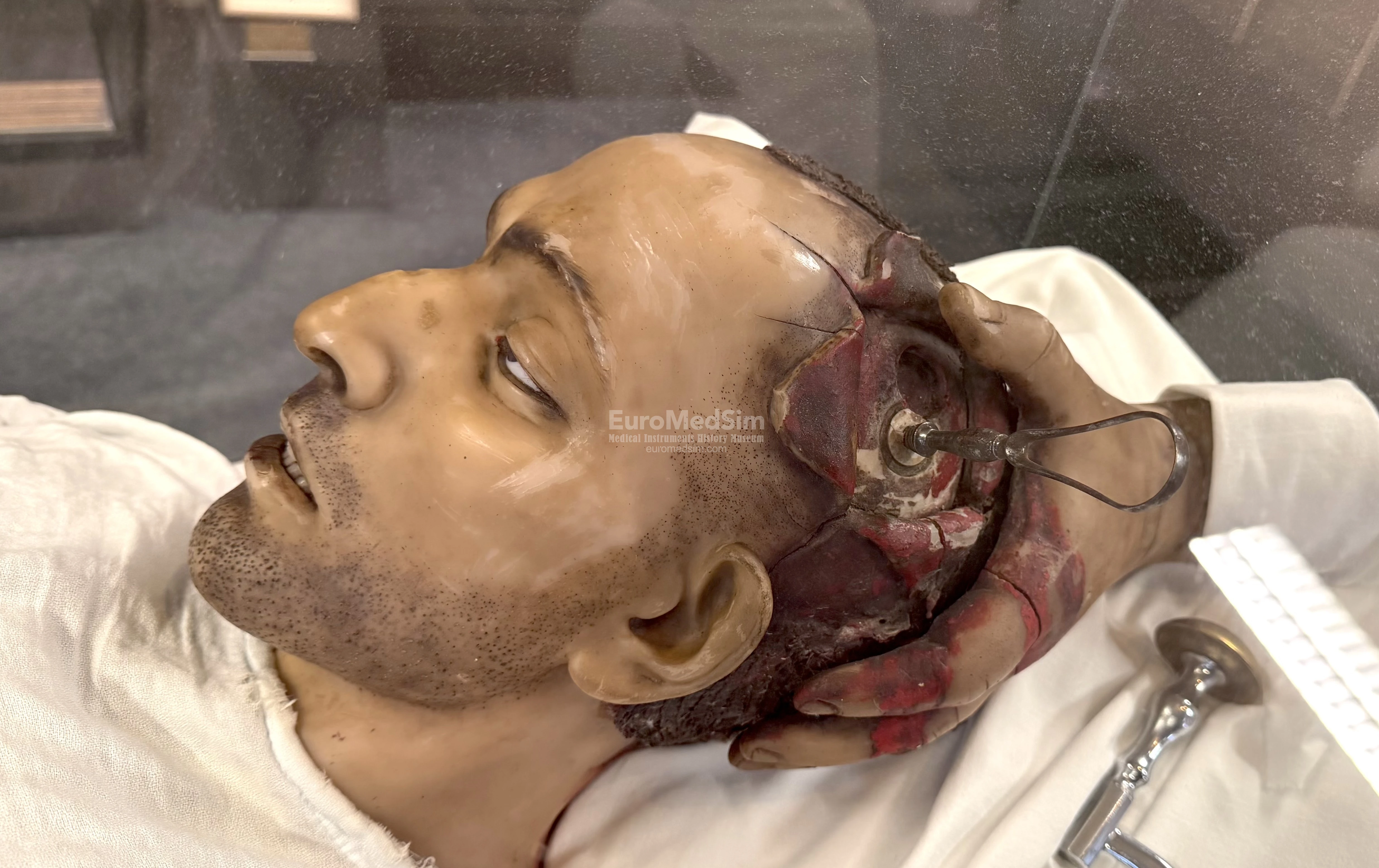
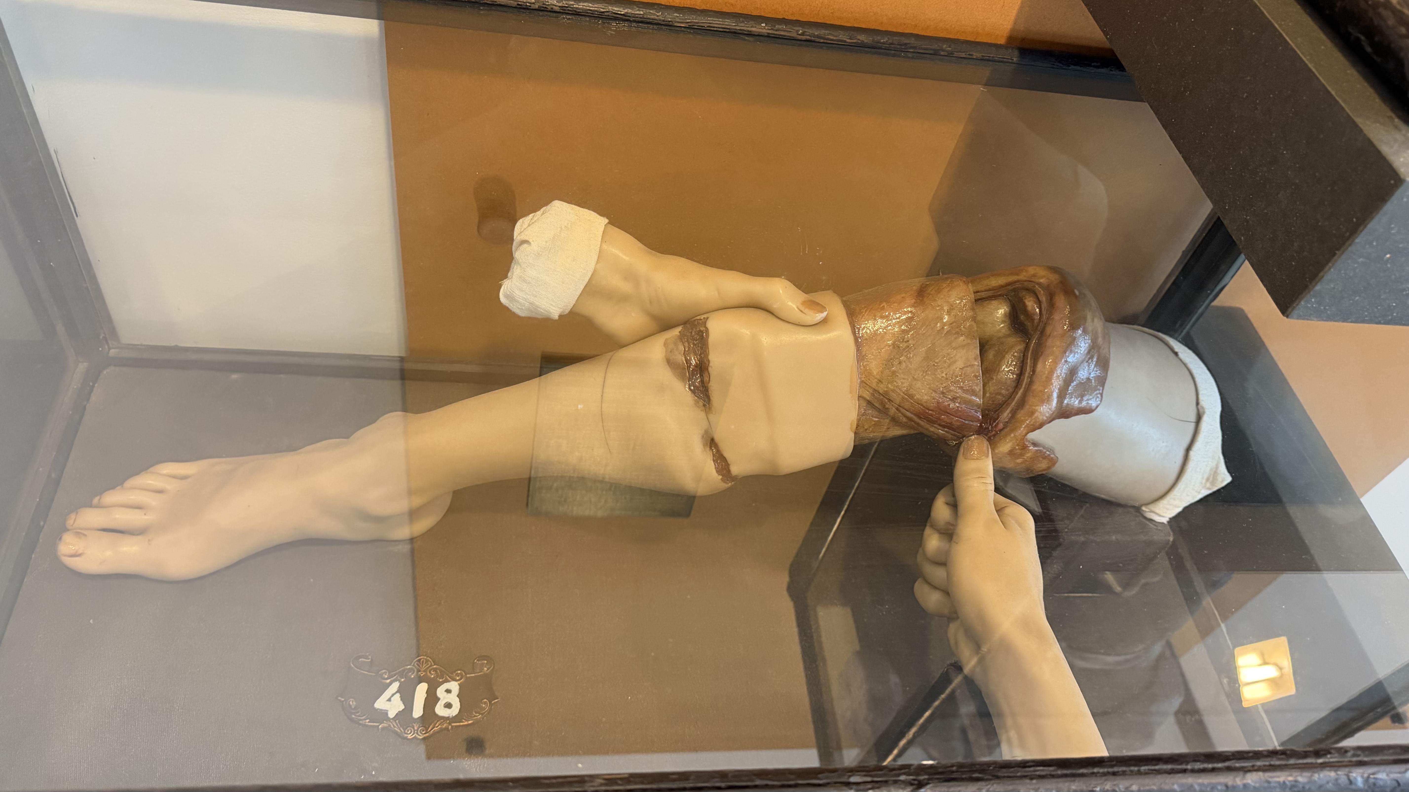
Trepanations were performed on patients suffering from skull fractures, convulsions, epilepsy, or psychiatric problems. The removed fragments were used as protective pendants. As the skull is relatively insensitive (unlike the scalp), this operation was relatively well tolerated. Cranial surgery was largely developed in the Middle Ages to remove the "stone of madness." Ambroise Paré used it to remove fragments of cranial fractures on the battlefield or to treat collapsed skulls. In the 19th century, it was used to drain abscesses. The kits shown include drill bits, files, lenticular knives, circular trepans, elevators, and so on. The anatomical wax in the center of the room represents a soldier wounded in the head during the Franco-Prussian War of 1870, on whom a trepanation is performed.
In the military context, amputations dominated surgery at the start of the 19th century. On the battlefield, more than 200 legs were amputated with saws in 24 hours (Dominique Larrey, Napoleon's surgeon, could detach a knee in less than 7 minutes) at the price of mortality reaching 30%, or even 85% for amputations above the knee. The cut was through the gangrenous part and, according to Paré, after ligation of blood vessels. From the 17th century, a tourniquet was used to compress part of the leg and prevent haemorrhage. Muscles were cut in strips rather than in a circle. Local care was limited, apart from cold-water dressings and cleaning wounds with permanganate. Anesthesia was summary apart from the use of opium. During the retreat from Russia, Larrey noted that severe cold diminished patients' pain, and this procedure would long be retained for amputation of gangrenous limbs.
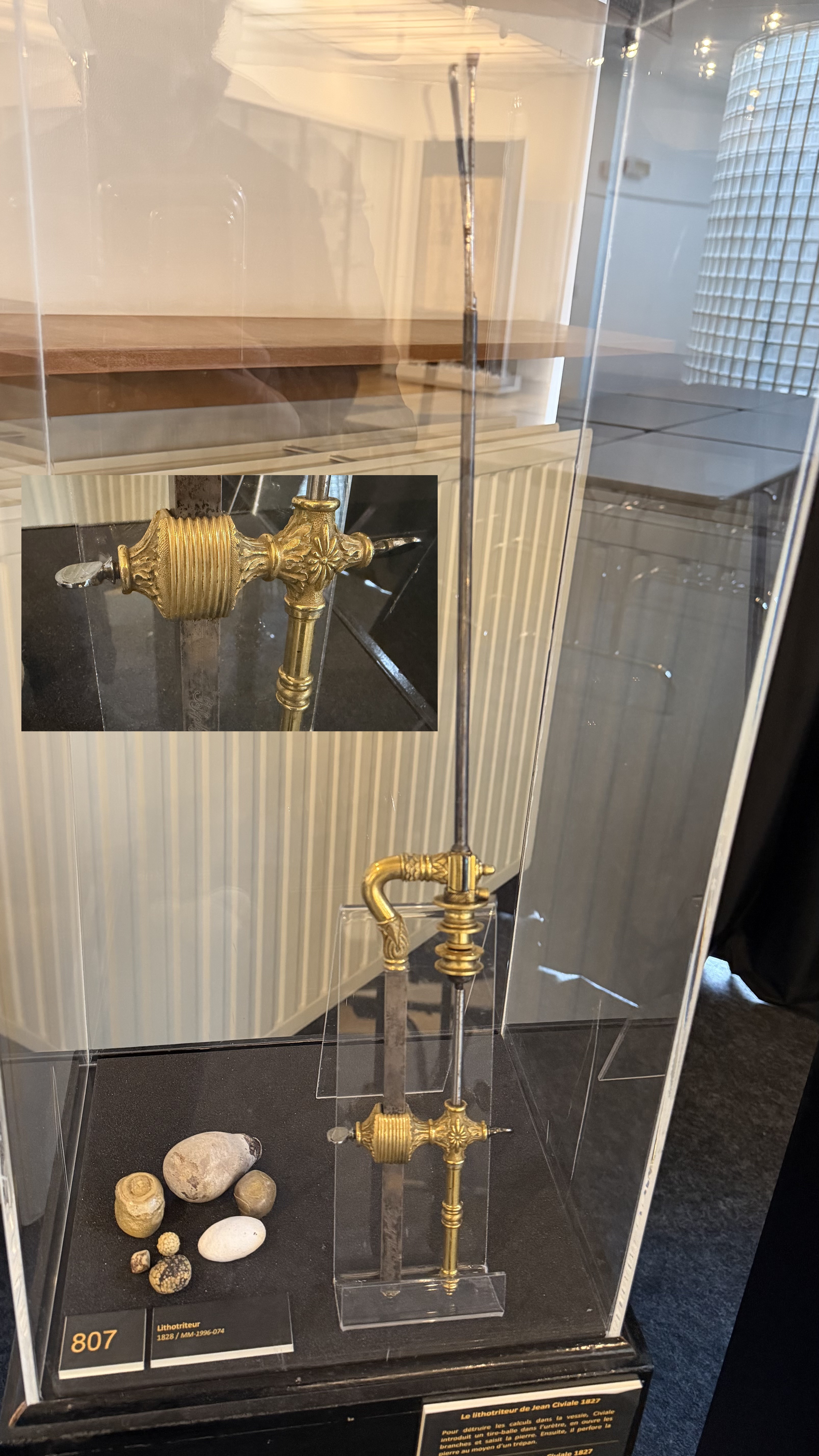
Over the centuries, bladder stones were one of the most frequent and painful conditions in men. In Antiquity, they were removed by excision, but this was not without risk. The stone was pushed to the anterior wall of the bladder by two fingers inserted in the rectum, and an incision was made over the bulge. Complications were frequent and results often disappointing, to the point that Hippocrates rejected it. The affirmation "I will not cut for stone" appears in his famous Oath—a phrase that for centuries influenced learned physicians who looked down on the barbers performing the operation. Vesical excision progressed little until it died out in favor of surgery through the urinary tract and pulverizing stones in the bladder. Jean Civiale popularized this method with his famous lithotrite.
Precolumbian Room: 2nd Floor
The Peruvians represented evident physical deformities on several terracottas, such as mouth ulcers that could be due to disease, a ritual process, or even criminal punishment. The appearance of the split lip curiously resembles the nostrils of the llama, a sacred animal sacrificed at ritual ceremonies. A so-called psychopomp llama was, according to beliefs, supposed to escort the dead to the afterlife. Amputations of the foot are also found. These remind us of the Creator god, who had a foot amputated, or indicate a more brutal practice of preventing a prisoner from escaping. In some tombs, amputees have even been found with leg prostheses to walk better.
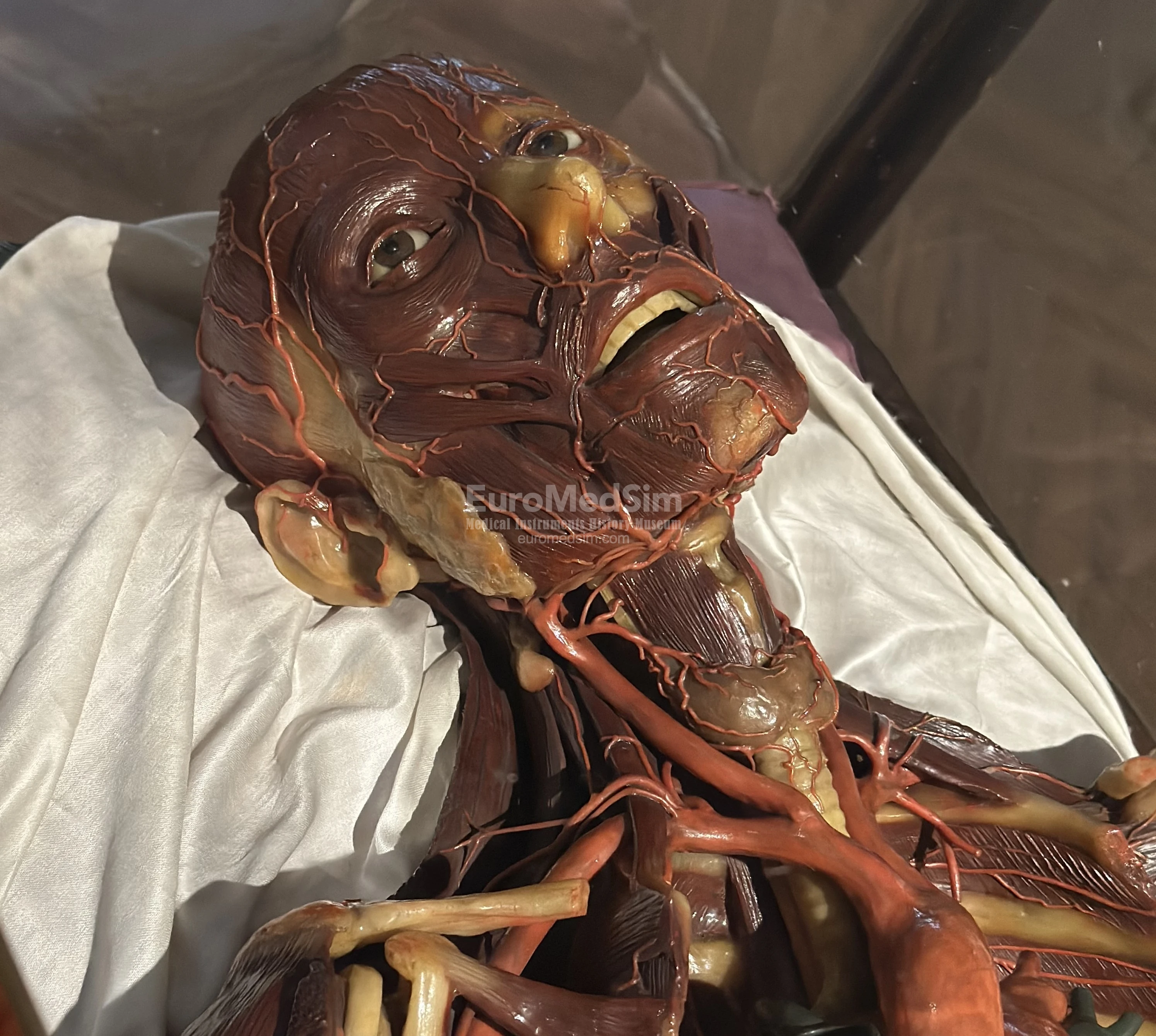
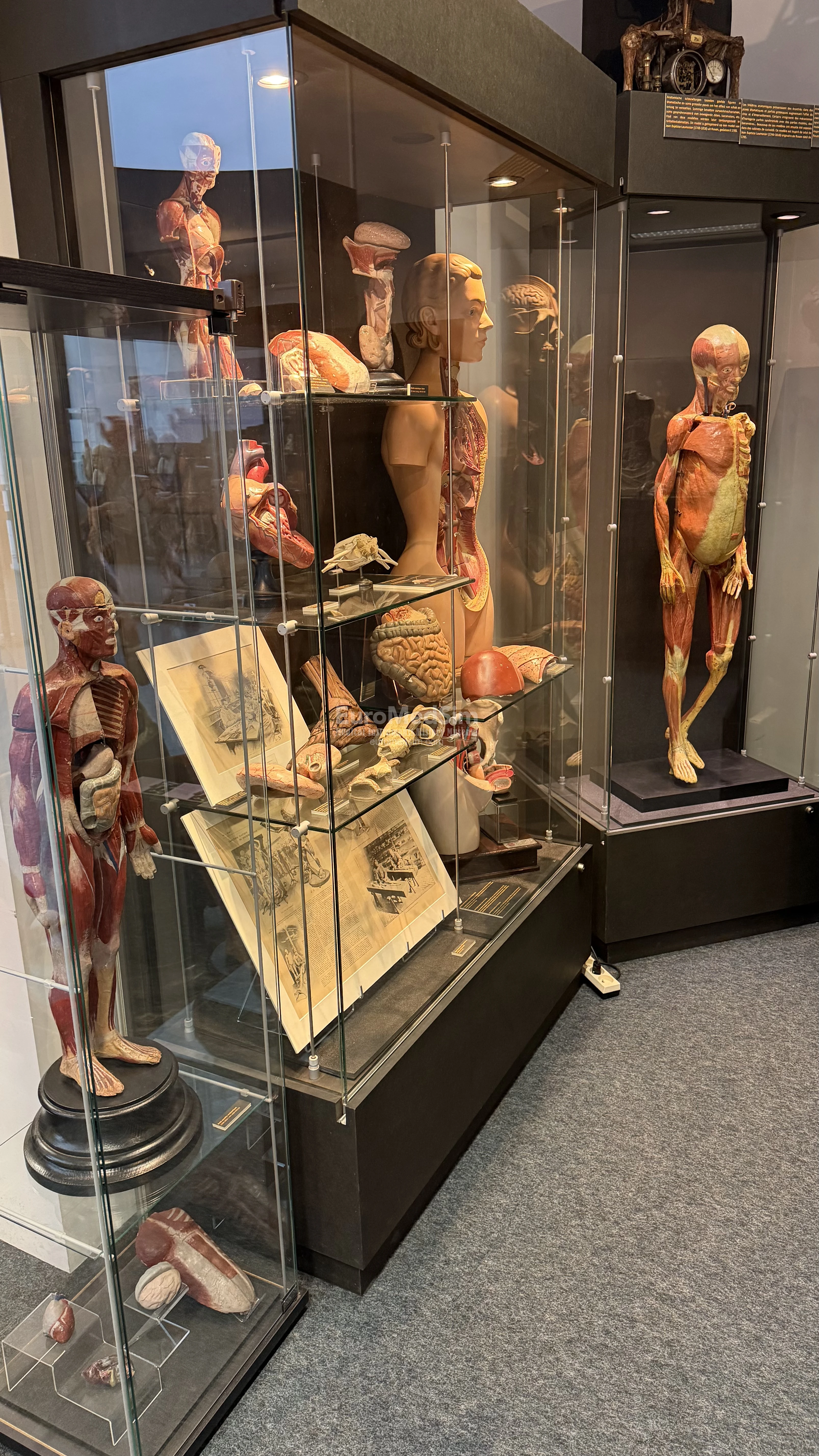
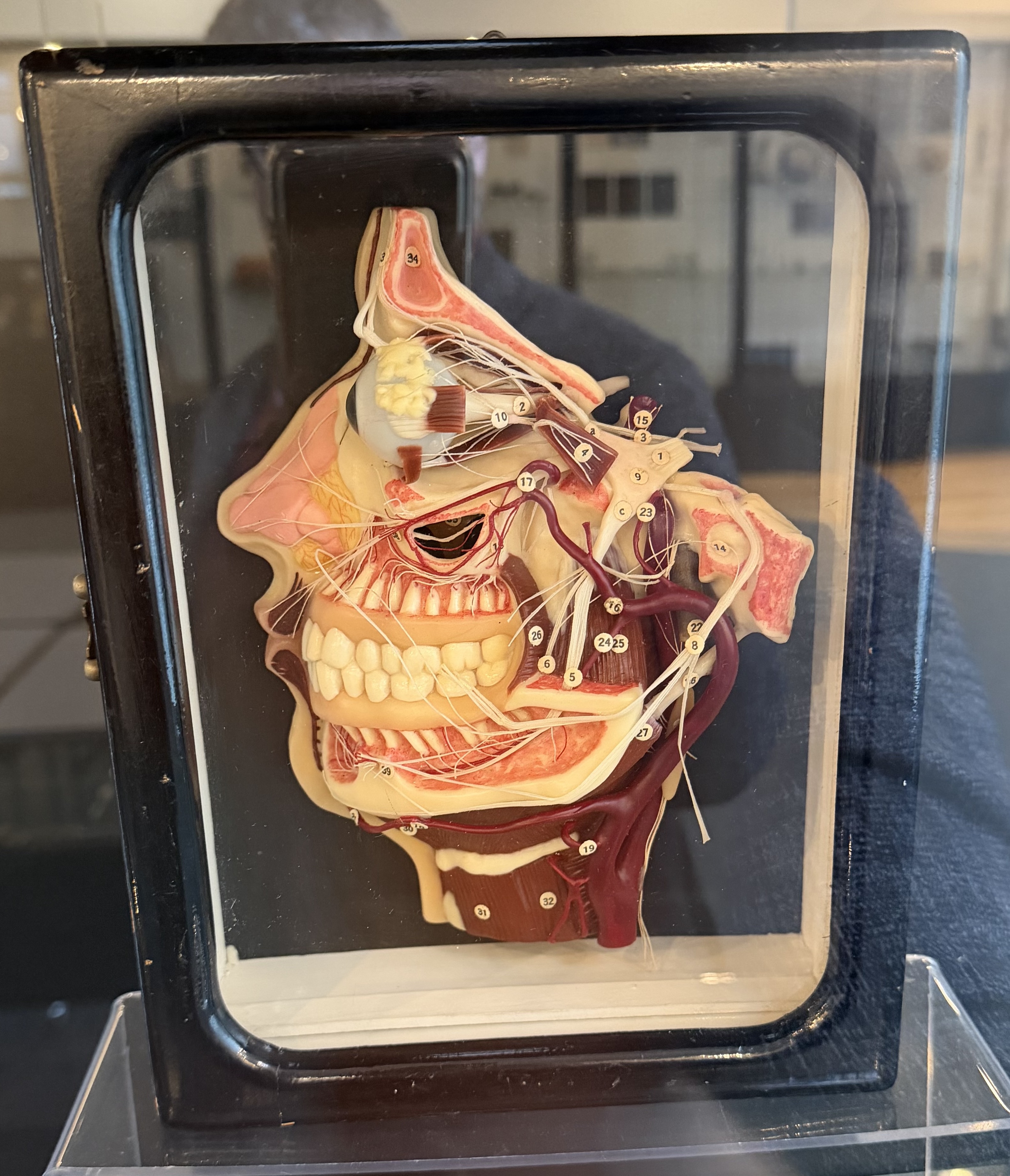
Wax anatomical and pathology models
The wax anatomical models displayed in this room show not only dissected bodies but also various surgical techniques—like amputations and the use of splints—alongside depictions of disease symptoms, embryonic development, and childbirth. Originally created in the early 1800s as teaching aids for medical students, they were later used to train paramedics and eventually became attractions at traveling fairs during the 20th century.
Inside the Spitzner Hell Room, visitors encounter wax figures that graphically illustrate the effects of syphilis, gonorrhea, alcoholism, and other conditions — sights that were common in the 1800s but have mostly vanished thanks to modern medicine. Although their lifelike detail was meant to educate, these models also functioned as a powerful deterrent, warning the public about the dangers of certain lifestyles.
Useful info
The Museum is situated next to the Erasme Hospital in Anderlecht (Brussels-Capital Region) and is accessible by car or public transportation in less than 30 minutes from the center of Brussels. The museum's address: 808 Route de Lennik, 1070 Anderlecht. Free admission with Brussels Card. Open 7 days a week (Working days 09:00–16:00, Sat/Sun 12:00–16:00, please check official website before visit).
Acknowledgements: the text on this page was compiled using materials provided on the official website and during a visit to the Museum of Medicine (Musée de la Médecine / Museum van Geneeskunde).